

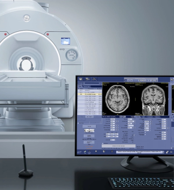
For neuroradiologists, the Signa Magnus 3T MRI system may offer enhanced visualization of the brain microstructure, microvasculature, and function.
Senior Editor, Diagnostic Imaging
For neuroradiologists, the Signa Magnus 3T MRI system may offer enhanced visualization of the brain microstructure, microvasculature, and function.

The presence of fat-enlarged axillary lymph nodes on mammography screening exams is associated with a fourfold higher risk of type 2 diabetes and a 2.6-fold higher risk for cardiovascular disease, according to new research presented at the ARRS Annual Meeting.
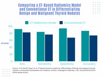
A computed tomography (CT)-based radiomics model that includes 28 radiomic features showed significantly higher accuracy, sensitivity, and specificity than conventional CT in differentiating benign and malignant thyroid nodules, according to newly published research.
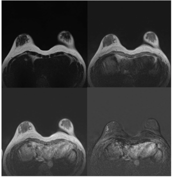
Supplemental breast MRI had a cancer detection rate (CDR) of 20/1000 and a positive predictive value (PPV) of 50 percent, according to preliminary findings from a prospective trial involving women with heterogeneously or very dense breasts.
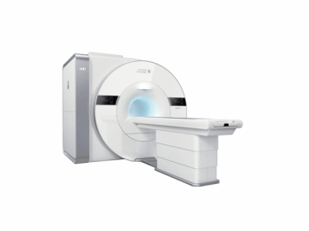
In addition to enhanced resolution, the uMR Jupiter 5T device reportedly addresses image uniformity and radiofrequency safety that have been previous challenges with ultra-high-field MRI.
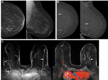
For over 1,200 women with a previous history of breast cancer, interim findings from a prospective trial suggest that adding contrast-enhanced mammography (CEM) to digital breast tomosynthesis (DBT) enhances detection of second malignancies with a limited increase in recalls.
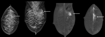
The 10-study meta-analysis demonstrated that contrast-enhanced mammography has a 95 percent sensitivity rate and an 81 percent specificity rate for diagnosing suspicious lesions in women with dense breasts.
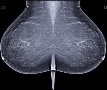
In finalized updates to breast cancer screening recommendations, the United States Preventive Services Task Force (USPSTF) advocates biennial mammography screening for women 40 to 74 years of age and notes insufficient evidence for the use of supplemental MRI in women with dense breasts.
In a recent interview, Steven Raman, M.D., discussed the use of MRI-guided transurethral ultrasound (TULSA) in the treatment of intermediate risk prostate cancer and key research findings with one- and five-year outcomes.
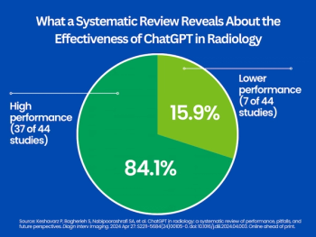
While noting a variety of pitfalls with the chatbot ranging from hallucinations to improper citations, the review authors found the use of ChatGPT in radiology demonstrated “high performance” in 37 out of 44 studies.
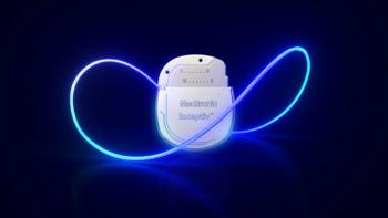
Reportedly the smallest and thinnest implantable spinal cord stimulator (SCS) device, the Inceptiv closed-loop SCS modality allows access to 1.5T and 3T full-body MRI without power restrictions.
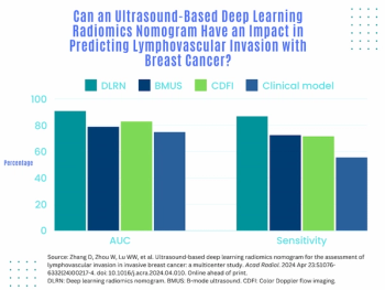
An emerging deep learning radiomics model, based on B-mode ultrasound and color Doppler flow imaging, demonstrated a 91 percent AUC for predicting lymphovascular invasion in a multicenter study of women with invasive breast cancer.

Combining a support crossing catheter with intravascular ultrasound imaging, the Provisio SLT IVUS System offers simultaneous real-time visualization and measurement of vessel lumen.
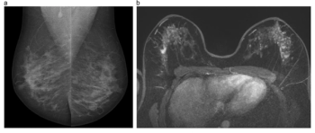
One of the recommendations from the European Society of Breast Imaging (EUSOBI) is annual breast MRI exams starting at 25 years of age for women deemed to be at high risk for breast cancer.
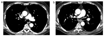
In comparison to energy-integrating detector CT for the workup of suspected acute pulmonary embolism, the use of photon-counting detector CT reduced radiation dosing by 48 percent, according to newly published research.
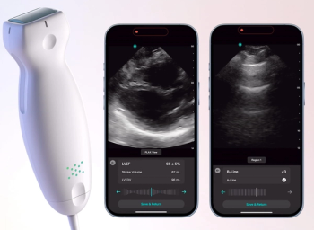
The artificial intelligence (AI)-powered applications reportedly allow clinicians to diagnose pulmonary edema and measure left ventricle ejection fraction within seconds.
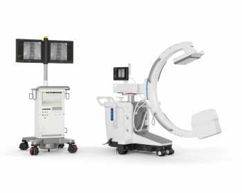
In addition to advanced imaging quality and dose efficiency, the Philips Zenition 30 mobile C-arm device emphasizes personalized user profiles and automated customization to help reduce procedure time.
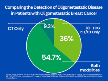
In a study of women with oligometastatic breast cancer, the use of 18F-FDG PET/CT detected additional metastases in one-third of cases that were not evident on conventional CT.
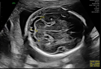
One of the features on the new Voluson Signature 20 and 18 ultrasound devices reportedly uses automated AI tools to facilitate a 40 percent reduction in the time it takes to perform second trimester exams.
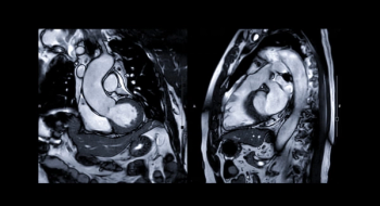
While noting inconsistencies with the diagnostic yield of cardiac MRI in patients who survived sudden cardiac arrest, researchers cited unique advantages in characterizing ischemic cardiomyopathy (ICM) and facilitating alternate diagnoses.
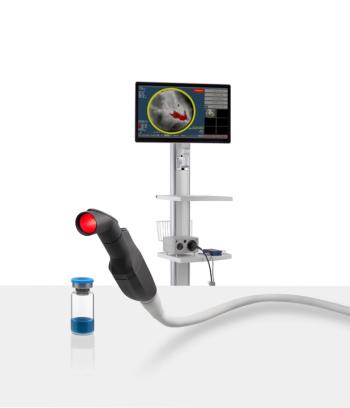
The combination of the optical imaging agent Lumisight and the fluorescence imaging device Lumicell Direct Visualization System, collectively known as LumiSystem, reportedly offers 84 percent accuracy with real-time detection of residual breast cancer after lumpectomy procedures.
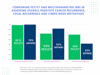
A recent meta-analysis found that multiparametric MRI and PET/CT had comparable detection rates for diagnosing prostate cancer recurrence.
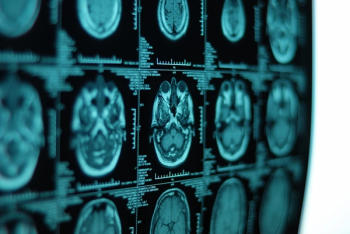
The use of continuous ofatumumab in patients within three years of a relapsing multiple sclerosis diagnosis led to substantial reductions in associated lesions on brain MRI scans, according to research recently presented at the American Academy of Neurology (AAN) conference.
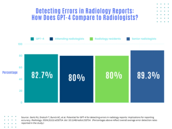
A new study shows that GPT-4 may offer comparable error detection rates to those of attendings and radiology residents in reviewing radiology reports, but researchers noted key caveats as well.
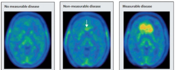
A positron emission tomography (PET) agent that targets LAT1 and LAT2 membrane transport proteins, TLX101-CDx or 18F-floretyrosine could be utilized to help characterize progressive or recurrent glioma.
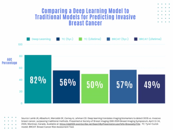
Artificial intelligence (AI) assessment of mammography images may significantly enhance the prediction of invasive breast cancer and ductal carcinoma in situ (DCIS) in women with breast cancer, according to new research presented at the Society for Breast Imaging (SBI) conference.
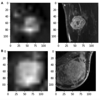
For the prediction of axillary lymph node metastasis in patients with breast cancer, an MRI-based, 4D convolutional neural network model demonstrated an AUC of 87 percent and sensitivity of 89 percent, according to new research.
Discussing findings from a new study presented at the Society for Breast Imaging (SBI) conference, Shahrzad Tavana, M.D., detailed the significant impact of training sessions for MRI technologists in improving breast positioning, optimal field of view and accuracy of sequence submissions to PACS for breast MRI exams.
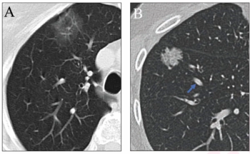
The inclusion of simulated adjudication for resolving discordant nodule classifications in a deep learning model for assessing lung adenocarcinoma on chest CT resulted in a 12 percent increase in sensitivity rate.
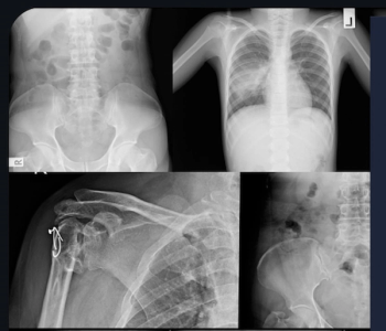
Leveraging artificial intelligence, the Rho software assessment of X-rays may allow earlier detection of bone loss.