The ViT model provided the highest specificity rate at 70.5 percent in comparison to 60.9 percent for the radiomics model and 60 percent for the combined model, according to the researchers.
“The Radiomics-ViT model demonstrated the highest sensitivity in detecting VPI, suggesting its potential to support more aggressive treatment strategies, particularly for younger patients with good pulmonary reserve. Although this approach may entail a certain risk of false positives, the overall assessment prioritizes minimizing the risk of undertreatment and potential recurrence,” noted lead study author Yuanxin Sun, MD, who is affiliated with the Department of Radiology at the Shanghai Chest Hospital and the Shanghai Jiao Tong University School of Medicine in Shanghai, China, and colleagues.
Three Key Takeaways
• Combined radiomics/ViT model maximizes sensitivity for VPI detection. In external testing, the combined model achieved 95.4 percent sensitivity for predicting visceral pleural invasion in LUAD ≤30 mm, outperforming radiomics alone (90.0 percent) and ViT alone (72.7 percent). This higher sensitivity comes at the cost of specificity (60 percent, lowest of the three models), so the model favors catching true VPI cases over minimizing false positives, a tradeoff the authors argue is appropriate to avoid under-treatment.
• This sensitivity profile may help guide more aggressive surgical planning. Given the risk of recurrence associated with missed VPI, the authors suggest the combined model's high sensitivity could support decisions toward more aggressive resection strategies, particularly in younger patients with adequate pulmonary reserve who can tolerate more extensive surgery.
• Component-specific feature extraction (solid, ground-glass, peritumoral) drives predictive performance. The ViT model's value came largely from separately extracting and weighting features across distinct tumor components rather than treating the tumor as homogeneous — with attention maps highlighting interfaces between components as particularly informative regions.
What Model Features Fueled Predictive Power for Visceral Pleural Invasion?
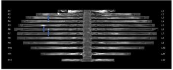
However, the study authors maintained that the ViT model, which was comprised of 12 sequential encoder layers with 12 attention heads, was critical for enhancing prognostic assessments for VPI in this patient population.
“ViT features, extracted from and aggregated across solid, ground glass, and peritumoral components, contributed most to VPI prediction. Separately extracting features from these regions allows the model to preserve component-specific information and prevents the dilution of biologically meaningful signals. Attention maps assigned higher weights to regions at some interfaces between different tumor components,” added Sun and colleagues.
(Editor’s note: For related content, see “Can Density Homogeneity on Chest CT Improve Differentiation of Sub-Centimeter Nodules?,” “Study Shows Merits of CT Vascular Sign for Differentiating Solid Pulmonary Nodules” and “Can AI-Enhanced Radiomics Improve Differentiation of Pulmonary Nodules on Chest CT?”)
In regard to study limitations, the authors acknowledged a lack of direct imaging and pathology registration to assess pathological characterizations emphasized by attention maps and conceded that the segmentation threshold was based on observational experience and prior literature. The researchers also noted that CT semantic features and clinical characteristics were not considered in the evaluation of the radiomic, deep learning and combined models in the study.