

Commentary|Videos|August 13, 2026
Breast Imaging in Focus: What New Research Reveals About Contrast-Enhanced Mammography in Prevalence and Incidence Screening Rounds
Author(s)Manisha Bahl, MD
In the latest episode of her “Breast Imaging in Focus” series, Manisha Bahl, MD, MPH, FSBI, offers her perspective on a new study that examines the effectiveness of contrast-enhanced mammography in initial and subsequent rounds of breast cancer screening.
Advertisement
Contrast-enhanced mammography (CEM) has received increasing attention as a supplemental screening tool for women at increased risk for breast cancer. CEM combines conventional low-energy mammographic images with recombined images obtained after administration of contrast material. The recombined images allow us to identify areas of enhancement related to tumor vascularity.
One of the important questions about any screening modality is whether its performance during the first or prevalence screening round is maintained with subsequent or incidence screening rounds. This distinction is important because prevalence screening tends to identify a reservoir of previously undetected cancers. Once those cancers are identified, cancer detection rates may decline with subsequent screening rounds.
Most studies evaluating CEM have focused on the first prevalence screening round. The purpose of a recent study was to compare the performance of CEM during prevalence and incidence screening in women at increased risk for breast cancer.1
The investigators conducted a retrospective single institution study of consecutive screening CEM exams performed between January 2015 and December 2021. Women undergoing CEM screening were typically at intermediate risk with an estimated lifetime breast cancer risk of 15 to 20 percent. Some women at high risk, defined as a lifetime risk of at least 20 percent, underwent CEM instead of MRI because of an MRI contraindication or personal preference.
The investigators classified an exam as a prevalence screen if the patient had no prior CEM and no breast MRI within the previous three years. An exam was considered an incidence screen if the patient had undergone prior CEM or breast MRI within the previous three years.
The final study population included 6,900 CEM screening exams in 2,700 women.Twenty-three percent of the exams were prevalent screens, and 77 percent were incidence screens.
The cancer detection rate was 19 cancers per 1,000 exams during prevalence screening compared with 11.6 cancers per 1,000 exams during incidence screening. Although numerically lower during incidence screening, this difference was not statistically significant after accounting for multiple exams in the same woman.
Importantly, sensitivity was maintained across screening rounds. Sensitivity was 86.1 percent for prevalence screening and 87.1 percent for incidence screening with no significant difference.
One of the most interesting findings relates to the added value of contrast enhancement. During the prevalence round, 60 percent of cancers were detected only because of enhancement on the recombined images and were occult on the low-energy mammographic images.
Importantly, this benefit persisted on subsequent screening rounds. During incidence screening, 58 percent of cancers were detected through enhancement alone. In other words, even after women had undergone prior vascular-based breast imaging, more than half of the cancers detected on subsequent CEM screening would not have been identified on the low-energy mammographic images alone.
Another important finding was the improvement in specificity. Specificity increased from 83.4 percent during prevalence screening to 91.5 percent during incidence screening, and this difference was statistically significant.
Overall, accuracy similarly increased from 83.5 percent to 91.4 percent. This finding is clinically relevant because one of the concerns with introducing a more sensitive screening modality is the potential increase in false positive findings and unnecessary biopsies. These results suggest that with subsequent CEM rounds and with prior vascular imaging available for comparison, CEM maintains its sensitivity while becoming more specific.
There are several limitations to keep in mind. This study was retrospective and performed at a single institution, which may limit generalizability. The patient population was also heterogeneous with respect to breast cancer risk. Although there was no statistically significant difference in cancer detection rates between prevalence and incidence screening, the authors note that the study was not sufficiently powered to detect differences in cancer detection rate.
The key finding is that the added value of CEM does not appear to be limited to the baseline screening exam. Contrast-enhanced mammography maintained high sensitivity with subsequent screening rounds, and approximately six in 10 cancers detected during prevalence and incidence screening were visible because of contrast enhancement. At the same time, specificity improved with incidence screening. These findings add to the growing evidence supporting CEM as a potential screening option for women at increased risk of breast cancer.
Dr. Bahl is an associate professor at Harvard Medical School and a breast radiologist affiliated with Massachusetts General Hospital.
Reference
- Lee CH, Amir T, Sevilimedu V, et al. Contrast-enhanced mammography for breast cancer screening in women at increased risk: comparison of prevalence and incidence screens. Radiology. 2026;320(1):e252433. doi: 10.1148/radiol.252433.
Advertisement
Related to this article

In the third of a three-part podcast episode, Wendie Berg, MD, PhD, FACR, FSBI, Manisha Bahl, MD, and Amy Patel, MD discuss recent developments with autonomous AI in breast imaging and share their perspective on the overall potential of AI to help mitigate the radiologist shortage.
In the second of a three-part podcast episode, Wendie Berg, MD, PhD, FACR, FSBI, Manisha Bahl, MD, and Amy Patel, MD discuss how to handle disagreements with adjunctive AI in breast imaging, whether to include AI assessments in PACS, and whether patients should pay for the use of adjunctive AI.
In the first of a three-part podcast episode, Wendie Berg, MD, PhD, FACR, FSBI, Manisha Bahl, MD, and Amy Patel, MD share insights on the utility of AI in breast imaging with respect to potential advantages, current concerns and future directions.

Catch up on the top radiology content of the past week.
In a recent interview, Amir Iravani, MD, FRACP, discussed the radiotherapeutic agent 177Lu-edotreotide and new dosimetry data from the COMPETE trial that was recently presented at the ASTRO conference.
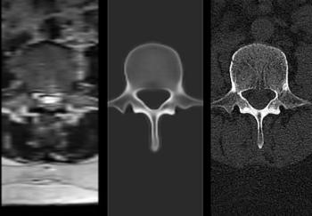
The cloud-based NextMR platform reportedly provides automated 3D bone images of conventional spine MRI images in less than 60 seconds.
In the latest episode of his “Interventional Radiology in Focus” series, Mina Makary, MD, discusses the utility of prostatic artery embolization for treating benign prostatic hyperplasia, key imaging considerations and pertinent technique pearls to help facilitate optimal outcomes.
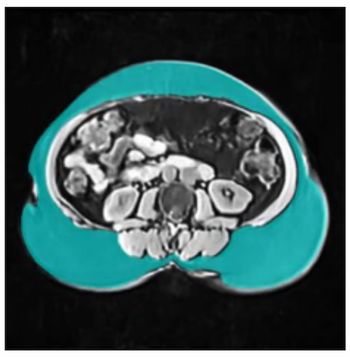
The AI-powered SwiftSight Body Composition software reportedly provides automated quantification of muscle and adipose tissue after a brief MRI scan.
Advertisement
Advertisement
Trending on Diagnostic Imaging
1
Key Sessions of Interest for Radiologists at the Upcoming HLTH USA Conference
2
The Reading Room Podcast: Current Perspectives, Challenges and Possibilities with AI in Breast Imaging, Part 3
3
Why Dutifully Waiting in Line is No Longer an Option for Advancement in Radiology
4
What New Dosimetry Data Reveals About 177Lu-Edotreotide for GEP-NET Treatment
5