The study authors also determined that DBT offered 37 percent higher detection of lobular carcinoma in situ, 45 percent higher detection of atypical lobular hyperplasia and more than double the detection rate for radial sclerosing lesions.
“ … Over nearly 2 decades, digital breast tomosynthesis was associated with increased detection of certain high-risk benign breast diseases at screening mammography, particularly radial sclerosing lesions,” noted lead study author Brian L. Sprague, PhD, who is affiliated with the Department of Surgery at the University of Vermont in Burlington, Vt., and colleagues.
What Were Possible Contributing Factors to the Increased Detection of Radial Sclerosing Lesions with DBT?
The doubling of detection for radial sclerosing lesions affirms previous study findings showing an increase of these cases after increased dissemination of DBT, according to the researchers.
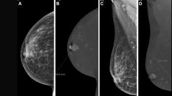
“This is likely due to the improved visualization of architectural distortion, which is a hallmark of radial sclerosing lesions. Changes in pathologist diagnosis of radial sclerosing lesions could also have contributed to the observed temporal trends,” suggested Sprague and colleagues.
The researchers noted annual percentage increases of 5.4 percent for radial sclerosing lesions and 19 percent for atypical papillary lesions. Other study findings revealed an annual percentage decline of 7.4 percent with cases involving atypical ductal hyperplasia (ADH) between 2009 and 2017 after a previous four-year period of 9.4 percent annual increases of ADH presentations.
Three Key Takeaways
• DBT finds more high-risk BBD than digital mammography. In this 17-year analysis of nearly 4.5 million screening mammograms, DBT had a 36 percent higher overall detection rate for high-risk benign breast disease. Detection was 37 percent higher for lobular carcinoma in situ, 45 percent higher for atypical lobular hyperplasia, and more than double for radial sclerosing lesions.
• The gain is largest for radial sclerosing lesions, likely because of architectural distortion. DBT shows architectural distortion, a hallmark of radial sclerosing lesions, more clearly. Radiologists reading DBT can expect to see these lesions more often, which fits with their 5.4 percent annual increase over the study period.
• Finding more high-risk BBD does not have to mean more surgery. In the accompanying editorial, Regina Hooley, MD, noted that management of radiology-pathology concordant radial sclerosing lesions, lobular carcinoma in situ, and atypical lobular hyperplasia has de-escalated over time. Many of these lesions can now be managed without automatic excision, which may ease concerns about overtreatment.
What About Possible Overtreatment with High-Risk Benign Breast Disease?
In an accompanying editorial, Regina Hooley, MD, a professor of radiology and biomedical imaging at the Yale School of Medicine, addressed concerns about overtreatment with high-risk BBD.
"Critics may argue that finding additional high-risk BBD requiring excision will lead to more unnecessary surgeries. However, while breast cancer screening has evolved, so has histologic diagnosis and management of high-risk BBD. Like for radial sclerosing lesions, management of radiology-pathology concordant lobular carcinoma in situ and atypical lobular hyperplasia has de-escalated over time," noted Dr. Hooley, a professor of radiology and biomedical imaging at the Yale School of Medicine.
(Editor’s note: For related content, see “Mammography-Based Deep Learning Model May Help Detect Precancerous Changes in High-Risk Women,” “Age and DBT Breast Cancer Screening: What the Research Reveals” and “Assessing the Impact of AI-Powered Slab Reconstruction on Reading Time for Digital Breast Tomosynthesis.”)
In regard to study limitations, the authors conceded that the observed temporal trends may not reflect changes in the incidence of different benign breast diseases, citing possible contributing factors such as changes with mammographic modalities, biopsy practices and classification schemes over time.