


New research shows that higher total lesion volume in the brain for former football players is directly associated with higher vascular risks and changes in white matter hyperintensity-associated biomarkers of Alzheimer’s disease.
New research shows that higher total lesion volume in the brain for former football players is directly associated with higher vascular risks and changes in white matter hyperintensity-associated biomarkers of Alzheimer’s disease.
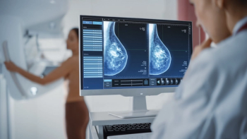
In a study examining the potential of the large language models ChatGPT-4 and Bard to follow ACR Appropriateness Criteria for breast cancer, lung cancer, ovarian cancer and colorectal cancer screening, researchers noted “impressive accuracy in making radiologic clinical decisions.”
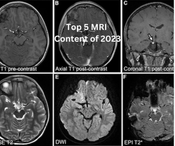
Catch up on the most well-read magnetic resonance imaging (MRI) articles from 2023.
Review the case study and test your knowledge to make the correct diagnosis.

Catch up on the top radiology content of the past week.
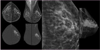
Contrast-enhanced mammography had a 98 percent sensitivity rate for diagnosing invasive lobular carcinoma and provided high conspicuity for 82 percent of detected lesions, according to research presented at the recent Radiological Society of North America (RSNA) conference.
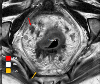
In one of the study periods from recently published research on rectal MRI acquisition, researchers found that 39.7 percent of rectal MRI exams adhered to guidelines from the Society of Abdominal Radiology (SAR) and only 6.8 percent followed guidelines from the European Society of Gastrointestinal and Abdominal Radiology (ESGAR).
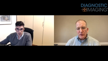
In a recent interview, Alexander Rau, M.D., discussed emerging research, presented at the recent RSNA conference, that shows the capability of diffusion microstructural imaging to differentiate subtle shifts in microstructural gray matter associated with common symptomatology of long Covid.
Catch up on the top radiology content of the past week.

In a recent interview, Mahsa Dolatshahi, M.D., M.P.H., and Cyrus A. Raji, M.D., Ph.D., discussed MRI and PET study findings, presented at the Radiological Society of North America (RSNA) conference, that showed an association between higher amyloid PET tracer uptake in the precuneus cortex and a higher visceral to subcutaneous fat ratio.
Catch up on the top radiology content of the past week.

Catch up on the top AI-related news and research in radiology over the past month.
BrainSpec Core reportedly offers enhanced sensitivity for low-grade gliomas and may facilitate the diagnosis of conditions including Alzheimer’s disease, multiple sclerosis, and epilepsy.
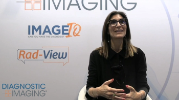
Dr. Kottler sat down with Diagnostic Imaging at RSNA 2023 to discuss AI imaging milestones and the potential impact of AI on workflows in radiology.
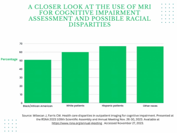
In a four-year study of over 1,600 patients who had outpatient head CTs, head CT angiography and/or brain MRI to assess cognitive impairment, researchers found that Black patients were over 9 percent less likely than White patients and over 16 percent less likely than Hispanic patients to receive brain MRI.
Catch up on the top radiology content of the past week.
Pixyl.Neuro reportedly leverages generative artificial intelligence (AI) technology to accelerate brain MRI assessment and improve early detection of abnormal atrophy.
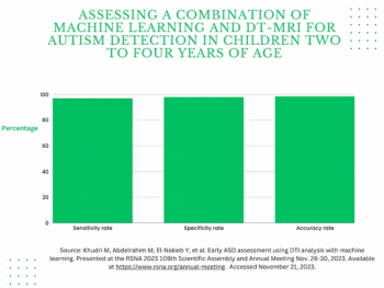
Through assessment of diffusion tensor MRI of the brain, a new AI system reportedly offers a 97 percent sensitivity rate in diagnosing autism in children between two to four years of age, according to research to be presented at the annual Radiological Society of North America (RSNA) conference next week.
Catch up on the top radiology content of the past week.
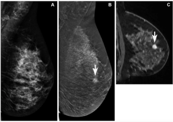
In findings from an enriched cohort of asymptomatic patients with screening-detected abnormalities, researchers found that contrast-enhanced mammography (CEM) was superior to conventional mammography and offered equivalent detection of breast cancer in comparison to breast MRI and abbreviated breast MRI.
Providing automated brain volume calculations based on MRI images, NeuroShield’s artificial intelligence (AI)-powered technology may help facilitate treatment for neurodegenerative conditions ranging from Alzheimer’s disease to epilepsy.
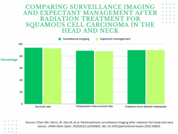
For patients who had a complete metabolic response to radiation treatment for head and neck cancer, the authors of a recent study found that surveillance imaging with PET/CT, MRI or CT did not improve outcomes in comparison to expectant management.
Catch up on the top radiology content of the past week.
Noting that initial imaging sensitivity is a key factor in cost-effectiveness for patients who do not require acute treatment for dizziness, researchers found that specialized MRI (including multiplanar high-resolution DWI) provided the most benefit in a comparative trial of neuroimaging modalities.
Emphasizing factors such as tumor size, serum neutrophil count and arterial phase hyperenhancement proportion on MRI, a new diagnostic model outperformed eight staging systems for predicting advanced recurrence of hepatocellular carcinoma after liver resection.