


The important role breast ultrasound plays in the emergency department-and the improvements that need to happen.
The important role breast ultrasound plays in the emergency department-and the improvements that need to happen.
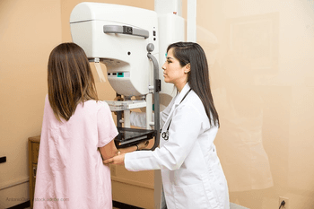
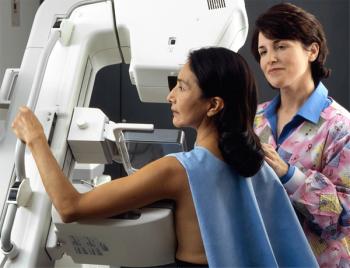
With new laws on the horizon, why it’s vital for radiologists to stay on top of the latest dense breast reporting information.

Practitioners are using the technology to make more accurate diagnoses.
Why researchers say practitioners and patients alike will be moving toward ultrasound-guided breast biopsies.

New laws and research are changing how dense breast notifications are happening.

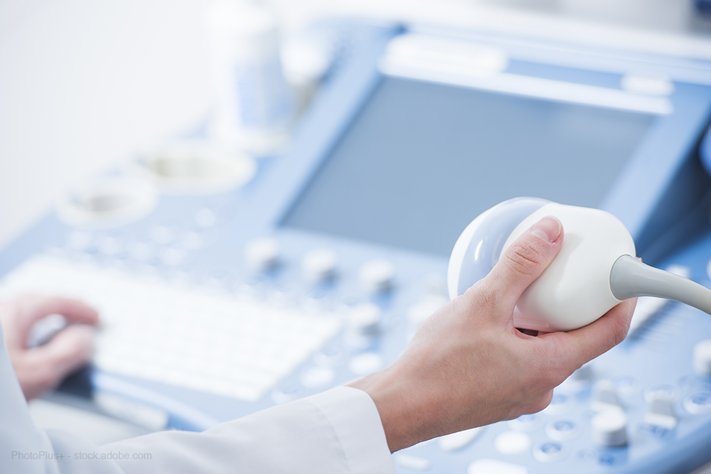
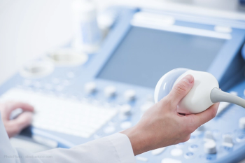
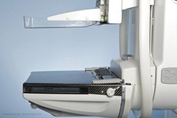
The market for automated breast ultrasounds is increasing. Here’s why.

Women who participated in regular breast screening programs benefitted more from therapy than women who were not screened regularly.
Birth defects can be tough to spot on ultrasounds, but new technological advances could make it easier.
Not following ACR practice guidelines potentially leads to unnecessary workups and extra health care costs.
Algorithm may help better predict how likely it is a woman will have cancer in her future.
Biggest obstacle in ultrasound training is lack of dedicated faculty time.
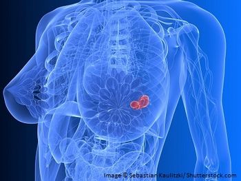
The problem of density and what it has meant for radiologists.

A total of 35 percent of cancers diagnosed after second-opinion review were not initially detected in the original interpretation.
Follow-up procedures are ordered by clinicians largely because current tools often cannot provide diagnostic certainty in identifying cancerous breast masses.
Imaging tests overused to diagnose pure breast pain among women.
Breast density legislation is still causing confusion and controversy among radiologists.
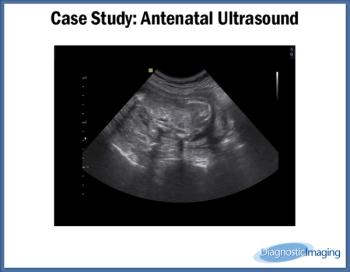
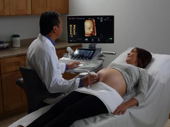
Case History: 25-year-old patient presents for antenatal ultrasound.
Ultrasound remains most sensitive imaging modality when diagnosing placenta accreta.
Ultrasound imaging used to identify fetal sex without a medical indication is becoming a public health issue and should be discouraged.
New diagnostic thresholds for ultrasound use in pregnancy may help avoid inadvertent harm to fetus.
Use of transvaginal sonography increases detection of ectopic pregnancies when patient history and clinical evaluation alone aren’t sufficient for diagnosis.

HealthDay News - Increased risks from diagnostic X-rays are slight, nonsignificant
Not only is it possible to detect aneuploidy and structural fetal anomalies with sonography during the first trimester, but doing so allows for better treatment options, according to a study published in the Journal of Ultrasound in Medicine.
A regional secretary of the Indian Radiology and Imaging Association and two other doctors were accused of conducting sex determination tests on decoy patients, according to an article in the Times of India.
Measuring the fetal zone of the adrenal gland is a better predictor of preterm birth than measuring cervical length, according to research presented at the Society For Maternal-Fetal Medicine Feb. 4.