


Late pregnancy scans cannot reliably predict one of the most common delivery complications.
Late pregnancy scans cannot reliably predict one of the most common delivery complications.

What to expect this week on Diagnostic Imaging.

Standard image-guided, vacuum-assisted biopsy can accurately identify remaining cancers in some patients with a less than 5 percent false negative rate.

Identifying a patient’s preferred method of communication can open the door for optimal, respectful patient care.
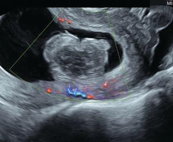
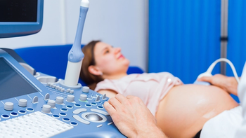
Ultrasound technology can be an indispensable tool for the management of suspected early pregnancy loss.
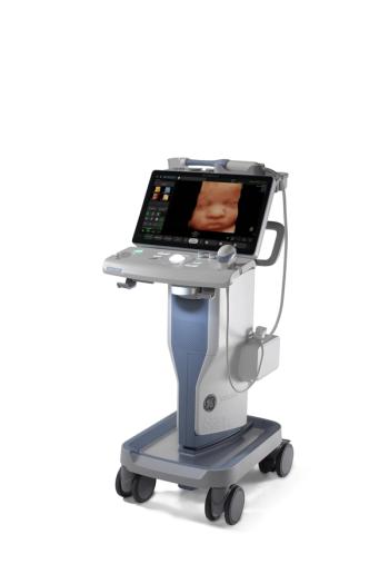
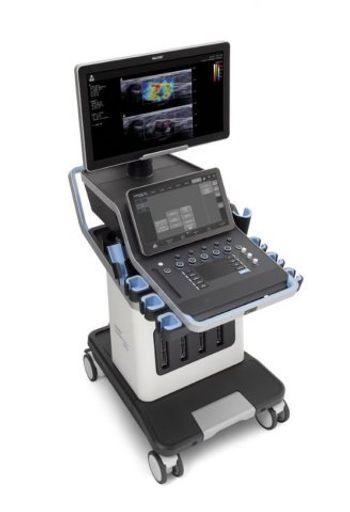
Voluson SWIFT is designed to shorten scan time and improve efficiency.

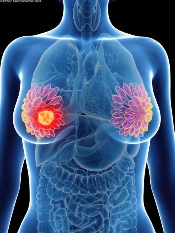
Breast imagers could be among the most highly burned out sub-specialists, but there are ways to reduce the work fatigue.
Radiation, alongside age and socioeconomic status, is associated with longer diagnosis-to-treatment timelines.

Rethinking CMR during COVID-19; Abdominal Imaging and COVID-19; Mental Health Impacts of COVID-19; African American and Lung Cancer Screening; Plus, African American Women and Disadvantages in Breast Cancer Screening
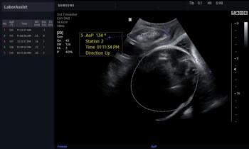
Ultrasound system provides obstetric measurements during labor in seconds, eliminating the need for digital vaginal exams and helping to side-step C-sections.

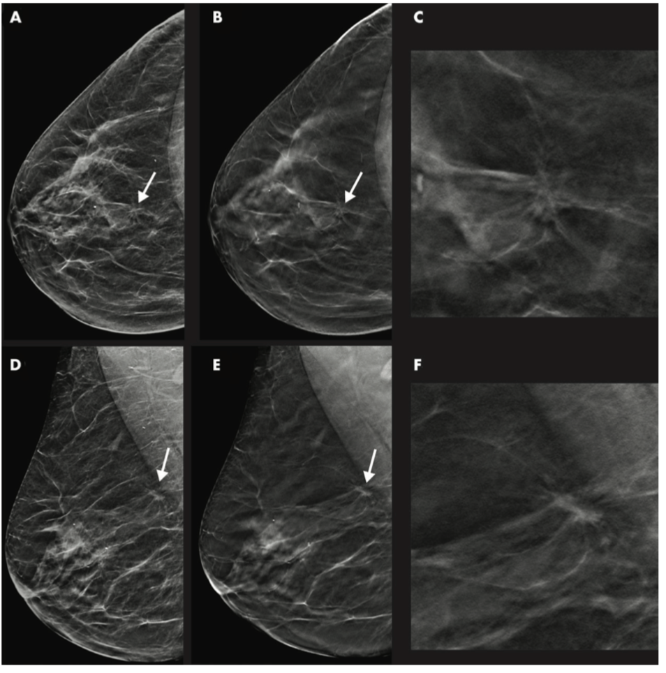
Ultrasound is an effective follow-up strategy for masses pinpointed with DBT, potentially side-stepping the need for digital mammography and lower radiation exposure.
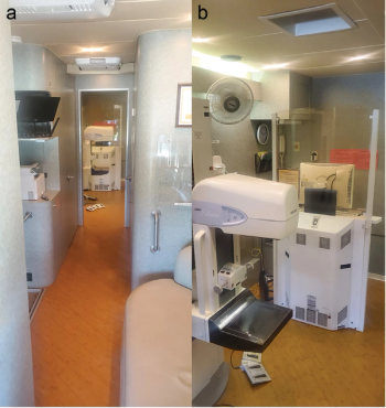
Houston-area mobile mammography program brings screening mammography and access to diagnostic services to women who have barriers to care.
The system is designed to reduce unnecessary biopsies, lesion correlation time, and improve overall diagnostic accuracy.

Breast cancers identified between patient screenings are more aggressive and lethal.

AI model can help alleviate increased sonographer workload, but still has high missed diagnosis rate.

After 2.5 hours, providers could successfully identify lungs with pathological patterns indicative of viral infection.

Eight recommendations released for imaging women post-mastectomy and breast reconstruction.
A third trimester ultrasound coupled with evaluating maternal characteristics could open door for monitoring and intervention.
Insights from industry experts on ways their departments have changed during the pandemic.

Study shows three risk factors for breast cancer can be passed down through genes.
Shortened protocol identifies more cancers than ultrasound or mammography.
Scans show no aggravation of symptoms.

Automated breast ultrasound overcomes limitations associated with hand-held technique.

Pediatric breast cancer rates are low, but guidelines exist to help alleviate patient concerns.

Why simulation-based training may be the best way to train residents.