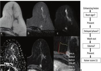
“Developing predictive criteria at baseline is critical for enabling personalized treatment strategies, as it allows oncologists to tailor treatment strategies before initiating therapy,” wrote lead study author Caroline Malhaire, M.D., who is associated with the Institute Curie and the Department of Medical Imaging at PSL Research University in Paris, France, and colleagues.
Overall, in the testing cohort, the multivariable model offered an 84.3 percent area under the receiver operating characteristic curve (AUC) for predicting post-NAT ARD. While acknowledging a 65 percent sensitivity rate, the study authors pointed out a 90 percent specificity and an 87 percent positive predictive value (PPV) with the multivariable model.
Three Key Takeaways
1. Baseline imaging predicts post-NAT ARD. Axillary ultrasound and breast MRI findings at baseline can help predict axillary residual disease (ARD) after neoadjuvant therapy (NAT), aiding personalized treatment strategies.
2. Key imaging markers for ARD risk. Patients with cortical thickening greater than 7 mm on ultrasound have a more than sixfold increased risk of developing ARD, with mean lymph node metastases significantly larger than those with thinner cortices (3.87 mm vs. 1.28 mm). Additionally, an anterior tumor location is strongly associated with ARD, increasing the likelihood by 14.1 times. In contrast, intratumoral high-signal intensity on T2-weighted MRI appears to be a protective factor, reducing the likelihood of post-NAT ARD by over 83 percent.
3. Multivariable model shows high specificity. The model demonstrated an 84.3 percent AUC, 90 percent specificity, and 87 percent positive predictive value (PPV), indicating strong predictive accuracy despite a 65 percent sensitivity rate.
Citing high false-negative rates with axillary assessments conducted after the use of NAT, the researchers maintained that baseline imaging evaluation provides greater clarity in ascertaining risks for post-NAT axillary residual disease.
“Baseline imaging, by assessing the whole tumor, optimizes MRI’s ability to evaluate tumor characteristics. Post-NAT evaluations, in contrast, are limited by small residual foci and treatment-related changes, while baseline imaging supports tailored treatment planning,” added Malhaire and colleagues.
(Editor’s note: For related content, see “Surveillance Breast MRI Associated with Lower Risks of Advanced Second Breast Cancer,” “Study: Conventional Mammography ‘Likely’ Misses More than 50 Percent of Breast Cancer Diagnoses” and “Can an Emerging AI Model Enhance Early Breast Cancer Detection on MRI?”)
Beyond the inherent limitations of a single-center retrospective study, the authors acknowledged a small testing cohort that prohibited subgroup analysis by subtypes of breast cancer.