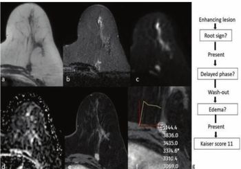
The study authors also noted that mpMRI offered a 89.4 percent positive predictive value (PPV) as well as a 97.9 negative predictive value (NPV).
“A high NPV (97.9%) suggests that low-risk NMEs can safely undergo short-term follow-up rather than immediate biopsy, potentially reducing unnecessary biopsies by approximately 14%,” posited Soliman and colleagues.
Three Key Takeaways
• mpMRI offers strong diagnostic performance for NME lesions. Consensus-based multiparametric MRI achieved 98.4 percent sensitivity, 86.4 percent specificity, and a 94 percent AUC when evaluating BI-RADS 3–5 non-mass enhancement lesions — outperforming any single imaging parameter alone. Combining DCE morphology, kinetics, and ADC measurements was key to this enhanced accuracy.
• A high NPV supports a "watch and wait" approach for low-risk NME. With a negative predictive value of 97.9 percent, mpMRI can help clinicians confidently defer immediate biopsy in favor of short-term follow-up for lesions that appear low-risk, with the authors estimating this could reduce unnecessary biopsies by approximately 14 percent.
• Specific MRI features are strong independent predictors of malignancy. Multivariable analysis identified several high-risk features clinicians should flag — most notably a type III washout curve (nearly 27× higher likelihood of malignancy) and segmental distribution (over 9× higher likelihood) — findings that can help prioritize which NME lesions warrant urgent workup.
Multivariable analysis revealed five MRI parameters that were statistically significant indicators of malignancy, including:
• a type II plateau curve associated with over a sixfold higher likelihood of malignancy;
• segmental distribution with over a ninefold higher likelihood of malignancy; and
• a type III washout curve, which had a nearly 27-fold higher likelihood of malignancy, according to the study authors.
(Editor’s note: For related content, see “Breast Radiologists Say New ACP Breast Cancer Screening Guidelines are ‘Many Steps Backward’ and ‘Dangerous’,” “New Breast MRI Study Says Background Parenchymal Enhancement Has No Impact on Lesion Detection” and “Breast MRI Study Links Peritumoral Edema to Reduced Disease-Free Survival from Breast Cancer in Young Women.”)
In regard to study limitations, the authors acknowledged the use of one 1.5T MRI system, the potential variability of values from ADC mapping across different field strengths and vendors, and the exclusion of BI-RADS 2 lesions. The study authors also noted that reviewing radiologists were blinded to patient clinical history.