

Blog|Articles|August 14, 2026
Could Photon-Counting CT Supplant Echocardiography for Primary Imaging of Transcatheter Heart Valves?
Author(s)Filippo Cademartiri, MD, PhD
The high-resolution temporal and spatial imaging with photon-counting CT bolster our understanding of the connections between anatomy and function in structural heart imaging.
Advertisement
For years, transcatheter heart valves have been assessed mainly by echocardiography with CT playing an important but often “structural” supporting role.
That paradigm is changing.
Photon-counting CT (PCCT) is not simply another incremental improvement in image quality. It is enabling a new level of morpho-functional characterization of transcatheter valves.
We can now visualize details that were previously blurred by blooming, noise, or limited spatial resolution. These details include:
• leaflet morphology with unprecedented definition;
• subtle leaflet thickening;
• hypoattenuated leaflet abnormalities (HALT);
• reduced leaflet motion;
• commissural configuration;
• frame expansion and deformation; and
• interaction with the native annulus and surrounding structures.
Perhaps even more importantly, high-resolution temporal and spatial imaging allows us to start connecting anatomy with function.
Why is one valve performing differently from another?Why do two apparently identical prostheses generate different gradients? How much of valve dysfunction is explained by geometry rather than biology?
These are no longer purely echocardiographic questions.
Photon-counting CT is evolving from a technology that “shows beautiful pictures” into one that explains why prosthetic valves behave the way they do. Morphology and function are finally being interpreted together rather than separately.
The future of structural heart imaging will not be defined by choosing between CT and echocardiography. It will be defined by integrating complementary information with PCCT providing anatomical insights that were previously inaccessible.
Seeing more is not the goal. Understanding more is.
Dr. Cademartiri is the director of advanced cardiovascular imaging and photon-counting CT at the Scientific Institute for Research, Hospitalization, and Healthcare Synlab Diagnostic Network in Naples, Italy. He is also a consultant in advanced cardiovascular imaging at CDI/Centro Diagnostico Italiano in Milan, Italy.
(Editor’s note: This blog is adapted with permission from Dr. Cademartiri’s original LinkedIn post at:
Advertisement
Related to this article

Does fractional flow reserve (FFR) or quantitative plaque burden from CCTA provide greater prognostic insight into coronary disease?
While better imaging may reduce the need for additional diagnostic tests, it may also eliminate the reimbursement that comes with those additional tests.

Catch up on the most well-viewed video interviews from Diagnostic Imaging in September 2026.

Catch up on the top radiology content of the past week.
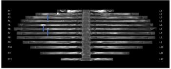
The use of adjunctive CT bone reading software in women with breast cancer detected between 8 to 22 percent more metastatic lesions for reviewing radiologists and reduced CT interpretation time by 50 percent or more, according to a new study.
Catch up on the top radiology content of the past week.

Catch up on key neuroradiology news and research from the past month, including recently published studies involving white matter hyperintensity findings on brain MRI, MRI-enhanced amyloid PET and the newly FDA-approved PET agent floretyrosine F18 for glioma imaging.

Magnetic resonance imaging offered a 29 percent higher sensitivity than CT for differentiating liver metastases, but CT provided 12 percent higher sensitivity than MRI for assessing vascular invasion, according to a new 40-study meta-analysis.
Advertisement
Advertisement
Trending on Diagnostic Imaging
1
Key Sessions of Interest for Radiologists at the Upcoming HLTH USA Conference
2
What New Dosimetry Data Reveals About 177Lu-Edotreotide for GEP-NET Treatment
3
The Reading Room Podcast: Current Perspectives, Challenges and Possibilities with AI in Breast Imaging, Part 3
4
Why Dutifully Waiting in Line is No Longer an Option for Advancement in Radiology
5