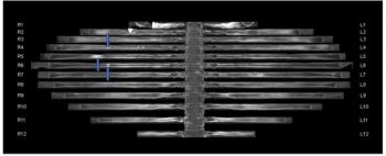
“Independence of margin status suggests that these imaging features do not predict a technically difficult resection at the SMA margin, but may instead suggest aggressive tumor or microscopic infiltration. Identifying these high-risk patterns preoperatively could prompt a shift in surgical management, potentially prioritizing these patients for neoadjuvant chemotherapy, although more studies are needed,” noted lead study author Kathy Truong, MD, who is affiliated with the University of Miami Miller School of Medicine, and colleagues.
The PPC2 pathway had significantly higher prevalence than the PPC1 pathway among patients with stage 2 and stage 3/4 disease in contrast to those with stage 1 disease, according to the researchers. The study authors noted that patients with the PPC2 pathway had a significantly lower median overall survival than those without the PPC2 spread (19.5 months vs. 51.4 months).
“The consistent association between PPC2 involvement on CT and adverse survival outcomes reinforces the role of PPC2 spread as a clinically meaningful imaging biomarker in PDAC,” added Truong and colleagues.
Three Key Takeaways
• The PPC2-pathway spread is a high-risk feature. In patients with EPNI on preoperative CT, involvement of the plexus pancreaticus capitalis 2 (PPC2) pathway was linked to more than a 2.5-fold higher mortality risk and a much shorter median overall survival (19.5 vs. 51.4 months). It was also more common in stage 2 and stage 3/4 disease.
• Morphology matters as much as location. EPNI with a mass-with-stranding pattern carried more than a 2.6-fold higher mortality risk. Median survival was 8.5 months with this pattern versus 48.8 months without it. Describing EPNI morphology may add prognostic value beyond anatomical extent.
• These features may change preoperative planning but the evidence is preliminary. Both findings predicted mortality independently of positive margin status. That suggests they reflect aggressive tumor biology or microscopic infiltration rather than a technically difficult resection. Flagging them before surgery could support prioritizing neoadjuvant chemotherapy. However, the data come from a small (n=89), single-center retrospective cohort with about 20 percent loss to follow-up so validation is needed before these potential markers guide management.
Patients who had a mass with standing morphology had over a fivefold lower median survival than those without it (8.5 months vs. 48.8 months), according to the study authors.
“These findings suggest that morphology may be as critical as anatomical location when assessing patient risk. In this context, we postulate that the mass with stranding pattern may reflect the anatomical manifestation of a more biologically aggressive or locally advanced tumor. While these observations are preliminary, they highlight the potential for refined morphologic assessment and risk stratification in PDAC,” emphasized Truong and colleagues.
(Editor’s note: For related content, see “MRI, CT or Ultrasound: Which Modality Offers the Most Utility for Imaging of Pancreatic Cancer?,” “MRI and CT Data Show Improved Progression-Free Survival with Emerging Agent for Gastroenteropancreatic Neuroendocrine Tumors” and “Can AI Enhance Non-Contrast and Contrast-Enhanced CT Detection of Pancreatic Cancer?’)
Beyond the inherent limitations of a single-center retrospective study, the researchers acknowledged the small size of the cohort, the loss of approximately 20 percent of the cohort to follow-up and the shorter median follow-up for those who EPNI versus patients who had EPNI.