


Opinion|Videos|May 1, 2026
Limitations of Conventional Imaging in Advanced Prostate Cancer
Author(s) Neal Shore, MD, FACS
Learn how next‑gen prostate cancer imaging outperforms CT and bone scans, revealing micrometastases and guiding team-based treatment decisions.
Advertisement
Episodes in this series

Welcome to the commentary with Dr. Neal Shore for the Diagnostic Imaging Peers and Perspectives series. In this episode titled, ‘Limitations of Conventional Imaging in Advanced Prostate Cancer’, Dr. Neal Shore highlights the limitations that have driven the need for more advanced imaging modalities. Over the past two decades, numerous phase 3 clinical trials evaluating therapies across multiple mechanisms of action have relied on conventional imaging techniques, including computed tomography (CT) scans and bone scans, and magnetic resonance imaging (MRI). These modalities formed the foundation for clinical trial endpoints, regulatory approvals, and guideline development across the FDA, EMA, and international regulatory bodies.
Despite their widespread use, conventional imaging approaches have notable limitations in sensitivity, particularly in detecting low-volume disease. Clinicians increasingly recognized that these tools often failed to identify disease in patients with rising prostate-specific antigen (PSA) levels, especially in those with biochemical recurrence or non-metastatic castration-resistant prostate cancer. Key patient populations, including those with negative conventional imaging but rising PSA following surgery, radiation, or androgen deprivation therapy, highlighted a significant unmet clinical need.
The discussion also references early guideline frameworks and index case analyses that emphasized these diagnostic gaps, particularly in patients with rapid PSA doubling times or biochemical relapse without visible metastases. These clinical scenarios underscored the importance of improving imaging sensitivity to better detect and stage disease and guide treatment decisions. Ultimately, the limitations of conventional imaging laid the groundwork for the development and adoption of more advanced imaging technologies aimed at improving detection, staging accuracy, and overall disease management in advanced prostate cancer.
In the next episode, ‘Multidisciplinary Interpretation of PSMA PET in Advanced Prostate Cancer,’ Dr. Neal Shore highlights the multidisciplinary approach required to interpret PSMA PET imaging and apply results to clinical decision-making in advanced prostate cancer. Effective collaboration and clear communication are essential to ensure accurate interpretation and optimal patient care.
Advertisement
Related to this article

Catch up on the latest imaging research presented at the recent American Society for Radiation Oncology (ASTRO) Annual Meeting.
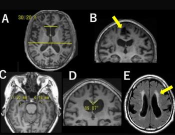
Tightness of the high-convexity sulci (THC) on brain MRI was associated with nearly threefold higher odds of cognitive decline over five to seven years of follow-up, according to a new prospective study involving over 2,100 community-dwelling senior citizens.
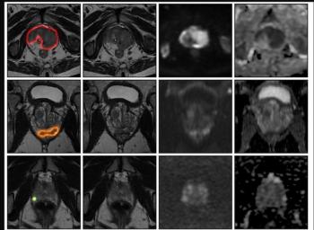
An AI model for prostate MRI offered an 89 percent AUC for prostate cancer detection in external validation testing, according to a newly published international multicenter study.

In the latest episode of her Breast Imaging in Focus series, Manisha Bahl, MD, MPH, FSBI, discusses new research comparing mammography-based AI models for breast cancer detection and short-term risk prediction.

In a recent episode of our Molecular Imaging in Focus series, Jack Andrews, MD, discussed pertinent findings from a recent intra-patient comparative study of PSMA PET agents in men with recurrent prostate cancer presented at the recent ASTRO conference.
The use of adjunctive CT bone reading software in women with breast cancer detected between 8 to 22 percent more metastatic lesions for reviewing radiologists and reduced CT interpretation time by 50 percent or more, according to a new study.
Expert Radiology is a **radiologist-run teleradiology practice with 10 years of experience**, hiring teleradiologists earning **$500–$900/hr with per-dictation pay**. Work from anywhere with true flexibility, **no shifts, no call, and no weekends**.
Our case mix is **predominantly outpatient and noncontrast**, with approximately **75% MRI**, plus X-ray and CT.
We cover **malpractice with tail**, handle all medical licensing, and ship you a **$10K reading station with six vertical 4K monitors**.
Minimum commitment is an **average of 400 cases per month**.
**Book a call with Brandi Escapule, Principal, Physician Services.**
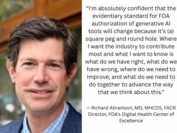
How will generative AI software be evaluated by the FDA in the future? What’s next for point-of-care ultrasound? What steps have been successful in facilitating adoption of AI applications across enterprise systems? Leaders in radiology and health care will be discussing these topics and more at the upcoming HLTH USA conference.
Advertisement
Advertisement
Trending on Diagnostic Imaging
1
What a Large Prostate MRI Study Reveals About AI and Unassisted Radiologist Assessment
2
MRI Study Links Tightness of High-Convexity Sulci to Higher Risk of Cognitive Decline in Older Adults
3
Breast Imaging in Focus: Can AI Models for Breast Cancer Detection and Risk Prediction Be Used Interchangeably?
4
Research Highlights from the ASTRO 2026 Annual Meeting
5