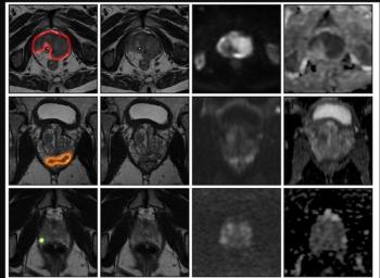
Employing a SUVmax threshold of 10, which corresponded to 100 percent specificity, the researchers utilized the combination of 18F-flotufolastat PET and MRI to classify findings as definitely positive, unclear and definitely negative groups. Accordingly, the study authors found that 22 patients with detected csPCa could proceed directly to definitive treatment without biopsy and suggested the omission of biopsy in 23 patients with a low risk of csPCa (a PI-RADS score < 2 and SUVmax < 3). In other words, combined 18F-flotufolastat PET/MRI may facilitate up to a 57 percent reduction of unnecessary prostate biopsies in comparison to a 25 percent reduction based on prostate MRI alone.
“18F-flotufolastat PET/MRI more than doubled the number of men with suspected prostate cancer for whom a biopsy may not be necessary to establish a diagnosis, compared with MRI alone. This high clinical impact was not due to an exceptional diagnostic accuracy of 18F-flotufolastat PET but rather to the combination of PI-RADS scores and 18F-flotufolastat PET SUVs, which allowed classification of the PET/MRI scans into 3 groups: definitely positive, definitely negative, and indeterminate,” noted lead study author Turkay Hekimsoy, MD, who is affiliated with the Department of Nuclear Medicine in the School of Medicine and Health at the Technical University of Munich in Munich, Germany, and colleagues.
Three Key Takeaways
• Combined 18F-flotufolastat PET/MRI substantially outperforms MRI alone for reducing unnecessary biopsies. Using a SUVmax threshold of 10 (100 percent specificity) alongside PI-RADS scoring, the approach enabled classification into definitely positive, indeterminate, and definitely negative groups, potentially sparing biopsy in up to 57 percent of patients, versus 25 percent with mpMRI alone.
• Diagnostic accuracy gains were modest and not statistically significant on their own. Combined PET/MRI showed numerically higher AUC (87.1 percent) than the PRIMARY score (86 percent), SUVmax alone (80.7 percent), or PI-RADS alone (75.2 percent), but the real clinical value came from the risk stratification framework (classifying scans into three groups) rather than superior discriminative accuracy per se.
• This should guide risk stratification, not replace biopsy outright. The study authors maintained that the 90 percent sensitivity rate obviously doesn’t omit potential cases of missed csPCa, and SUVmax can vary with reconstruction parameters and PSMA tracer choice. The researchers recommend applying this strategy selectively— in patients at very low or very high risk — rather than as a universal biopsy omission tool.
However, the researchers cautioned about potential variability with SUVmax, that may be affected by reconstruction parameters and the PSMA PET radiotracer one utilizes, and a 90 percent sensitivity rate that may not be high enough for completely safe omission of biopsy.
“Therefore, imaging-based strategies should be considered as a tool for risk stratification rather than a complete replacement of biopsy, with greatest relevance in carefully selected patients at very low or very high risk of csPCa,” advised Hekimsoy and colleagues.
(Editor’s note: For related content, see “Study: PSMA PET/CT Reduces Biopsy Rate by Nearly 50 Percent for Men with Equivocal or Non-Suspicious Prostate mpMRI,” “FDA Clears Emerging AI Software Application for Prostate MRI” and “Molecular Imaging in Focus: PSMA PET Radiotracers and Urinary Radioactivity: What Head-to-Head Prospective Multicenter Research Reveals.”)
Beyond the inherent limitations of a single-center retrospective study, the authors acknowledged the relatively high prevalence of csPCa in the cohort and that consensus image interpretation by experienced readers may have overestimated the impact of PSMA PET/MRI in real-world clinical settings with interpreting physicians of varying experience.