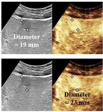
For patients with prior LR-4 MRI or CT assessments for lesions > 20 mm, the researchers found that CEUS had 100 percent specificity and PPV in LR-5 evaluations as well as a 71.9 percent accuracy rate. The study authors pointed out that CEUS led to management changes in 40 percent of these study participants.
Additionally, CEUS had 83.3 percent sensitivity overall and 85.7 percent sensitivity with LR-M categorizations of non-HCC malignancies in this patient population, according to the study authors.
Three Key Takeaways
1. CEUS provides higher specificity for LR-5 categorization. CEUS demonstrated 94.7 percent specificity and positive predictive value (PPV) in detecting HCC in patients with prior indeterminate MRI or CT LI-RADS LR-4 assessments, offering a significant advantage in identifying malignant liver lesions.
2. Management benefits for lesions > 20 mm. For liver lesions > 20 mm previously categorized as LI-RADS LR-4 on MRI or CT, CEUS achieved 100 percent specificity and PPV in LR-5 evaluations and resulted in management changes for 40 percent of patients.
3. Enhanced depiction of arterial phase hyperenhancement and washout. CEUS provides improved real-time sensitivity in detecting arterial phase hyperenhancement and allows for precise characterization of washout timing and degree. These features are particularly beneficial for the early diagnosis of HCC and distinguishing it from non-HCC malignancies.
Noting greater real-time sensitivity in showing arterial phase hyperenhancement in the liver with CEUS and the intravascular nature of ultrasound contrast agents in comparison to those employed for MRI and CT, the researchers said CEUS also provides more relevant insights into washout in the imaging of this patient population.
“ … CEUS has a unique property of allowing the detection of the timing of washout onset and characterization of the degree of washout, which is often beneficial for the early diagnosis of HCC and the differentiation of HCC from non-hepatocellular malignancies,” maintained Lyshchik and colleagues.
(Editor’s note: For related content, see “New CT and MRI Research Shows Link Between LR-M lesions and Rapid Progression of Early-Stage HCC,” “Can Intestinal Ultrasound Provide an Alternative for Evaluating Creeping Fat with Crohn’s Disease?”and “Can Photon-Counting CT Facilitate a Viable Alternative to MRI for Liver Fat Quantification in Patients with MASLD?”)
Beyond the inherent limitations of a retrospective study, the authors noted a relatively small cohort with the majority of the cohort having hepatitis C and metabolic dysfunction-associated steatohepatitis, limiting comparisons to other studies involving liver disease. The researchers also conceded that those who interpreted CEUS had access to patient CT and MRI findings.