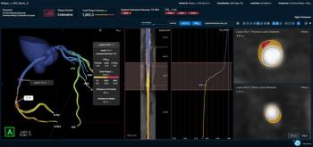
“In patients referred for ICA, CT-QFR yields a similar prognostic value as the combined assessment of ICA/SPECT. Given the noninvasive nature of CTA, CT-QFR may be an attractive alternative to nuclear stress testing and invasive examinations,” wrote lead study author Zehang Li, M.D., who is affiliated with the Department of Radiology at Ruijin Hospital and Shanghai Jiao Tong University in Shanghai, China, and colleagues.
“Particularly with respect to radiation exposure, the application of CT-QFR instead of ICA/SPECT could reduce the radiation dose. While CT-QFR is restricted to the evaluation of epicardial vasculature, SPECT is used to assess microvasculature perfusion, which can be affected by stenosis in epicardial arteries as well as microvascular dysfunction.”
The researchers noted that the median radiation dose for the ICA/SPECT combination was 21.7 mSv in comparison to 3.2 mSv for CTA.
Three Key Takeaways
1. Comparable prognostic value. CT-QFR provides a prognostic value similar to ICA/SPECT for predicting major adverse coronary events (MACEs) in patients with suspected or known coronary artery disease, making it a noninvasive alternative.
2. Radiation dose advantage. The median radiation dose of CT-QFR (3.2 mSv) reported in this study is significantly lower than that of ICA/SPECT (21.7 mSv), highlighting its potential benefit in reducing patient exposure.
3. Limitation with prior PCI patients. CT-QFR is less effective in predicting MACEs for patients with a history of prior percutaneous coronary intervention (PCI), due to imaging challenges such as blooming artifacts from implanted stents.
However, the study authors pointed out that CT-QFR is not as effective in predicting MACE for patients who have had prior percutaneous coronary intervention (PCI). In comparison to a 60 percent AUC for ICA/SPECT in this patient population, the researchers noted a 44 percent AUC for CT-QFR.
“Our study revealed a negatively influenced prognostic ability of CT-QFR in participants with a history of prior PCI. Previously implanted stents may generate blooming artifacts and result in segmentation error, posing challenges in distinguishing in-stent restenosis on CTA images,” noted Li and colleagues.
(Editor’s note: For related content, see “Assessing MACE Risk in Women: Can an Emerging Model with SPECT MRI Imaging Have an Impact?,” “Multicenter Study Shows Capability of AI CCTA Analysis for Predicting Major Adverse Cardiovascular Events” and “CT-Derived Fractional Flow Reserve Leads to Nearly 20 Percent Reduction of ICA in Cases of Suspected CAD.”)
In regard to study limitations, the authors noted that the high-risk cohort may preclude extrapolation of the study findings to a broader patient population. They also acknowledged that atheroma burden and atherosclerotic characteristics, which may enhance CTA-based risk stratification, were not evaluated in this study.