Nearly one out of every four patients with Long Covid (LC) had cardiac involvement findings on positron emission tomography/magnetic resonance imaging (PET/MRI) that were suggestive of myocarditis, according to a new prospective study.
For the study, recently published in the Journal of Nuclear Medicine, researchers examined imaging data for 98 patients (median age of 48.5) who had persistent cardiopulmonary symptoms nine to 12 months after initial COVID-19 infection. The study authors noted that 91 patients had PET/MRI scans, and 70 patients had dual-energy computed tomography (DECT). The study included a nine-patient control group who had a history of severe acute respiratory syndrome (SARS) with COVID-19 infection but did not have cardiopulmonary symptoms.
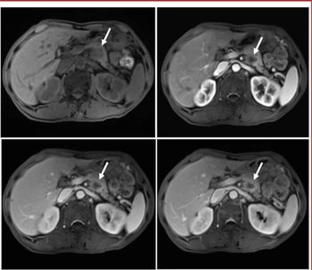
The study authors noted that 57 percent of patients had abnormal PET/MRI findings with 30 percent demonstrating aortic or pulmonary vascular uptake. In contrast to the control group that had no myocardial, pericardial or periannular uptake on PET/MRI scans, 24 percent of those with PET/MRI abnormalities had cardiac involvement suggestive of myocarditis and 22 percent had uptake suggesting possible pericarditis, according to the study authors. They added that 11 percent of the cohort had periannular uptake on PET/MRI scans.
“The imaging findings correlated with different serum biomarker profiles between LC and control subjects. This observation suggests that LC patients with long-term cardiopulmonary symptoms have a distinct plasma protein profile. Moreover, significant differences in plasma protein levels were observed between LC patients with normal and those with abnormal PET/MRI, supporting the hypothesis of specific humoral and immune mechanisms underlying pleiotrophic LC manifestation,” wrote lead study author Maria Giovanna Trivieri, M.D., an associate professor of cardiology, heart failure and cardiac transplantation with the Mount Sinai Health System in New York, and colleagues.