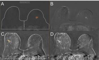
“The more pronounced improvements observed in our study may relate to the inherent advantages of multi-shot rs-EPI, including higher spatial resolution, reduced geometric distortion, and improved phase stability, which may provide a more stable foundation for DL-based reconstruction than ss-EPI (single-shot EPI),” noted lead study author Jin Joo Kim, MD, who is affiliated with the Department of Radiology with the Biomedical Research Institute and Pusan National University Hospital in Busan, Korea, and colleagues.
Achieving a Significant Reduction in MRI Scan Time with DLR Multi-Shot DWI
The researchers also noted these improvements with DLR multi-shot DWI came with a 29 percent reduction in MRI scan time (5.36 minutes vs. 7.52 minutes).
“This reduction was achieved through an optimized acquisition protocol combined with DL reconstruction, rather than fundamental shortening of the acquisition process itself. Conventional acceleration techniques, such as simultaneous multi-slice imaging, parallel imaging, and compressed sensing, have similarly reduced scan times while maintaining diagnostic quality. … Future studies integrating DL with such acceleration strategies could enable genuine scan time reductions in breast MRI workflows,” posited Kim and colleagues.
Three Key Takeaways
• Better image quality without added scan time. DLR multi-shot DWI significantly outperformed standard rs-EPI on key quality metrics — SNR, CNR, and lesion contrast — at both standard (b=800 s/mm2) and high (b=1500 s/mm2) b-values, while also cutting scan time by 29 percent (5.36 vs. 7.52 minutes). This suggests radiologists could gain sharper, more diagnostically useful DWI images without a workflow penalty.
2. ADC values appear reliable, but warrant validation. Although the primary cohort showed some differences in tumor and fibroglandular tissue ADC values between the two techniques, these differences didn't hold up consistently in the independent validation cohort, suggesting DL reconstruction may not introduce systematic bias in ADC quantification. Still, since ADC is used as a quantitative biomarker, sites should validate performance locally before relying on it clinically.
3. Scan time savings stem from reconstruction, not acquisition shortening
The time reduction came from an optimized acquisition protocol paired with DL reconstruction, not from fundamentally shortening the scan itself. The authors suggest combining DL with established acceleration techniques (parallel imaging, compressed sensing, simultaneous multi-slice) in future work could yield even greater efficiency gains.
Final Notes
While the primary cohort revealed lower tumor ADC values (0.97 x 10⁻³ mm2 vs. 1.02 x 10⁻³ mm2) and higher fibroglandular tissue ADC values (1.48 x 10⁻³ mm2 vs. 1.46 x 10⁻³ mm2) with DLR multi-shot DWI, the study authors said these values were comparable in independent validation testing.
“The absence of consistent ADC differences across cohorts suggests that DL reconstruction is unlikely to introduce systematic bias in diffusion quantification, although minor variations may occur. Previous studies have reported heterogeneous findings regarding ADC values, likely reflecting differences in DL algorithms, sequence types, and reconstruction strategies. Taken together, these findings underscore the importance of validating ADC performance when implementing DL-processed DWI in clinical practice, particularly when ADC serves as a quantitative imaging biomarker,” noted Kim and colleagues.
(Editor’s note: For related content, see “Can Multiparametric MRI Enhance Differentiation of Non-Mass Enhancement Breast Lesions?,” “New Breast MRI Study Saus Background Parenchymal Enhancement Has No Impact on Lesion Detection” and “Thirteen Takeaways on BI-RADS v2025 Changes for Mammography, Ultrasound and Breast MRI Reporting.”)
Beyond the inherent limitations of a single-center retrospective study, the authors acknowledged the relatively small size for the independent validation cohort, the use of one vendor for the deep learning algorithm and the lack of assessment for diagnostic performance.