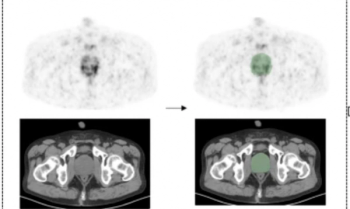
New research shows that 18F-NaF PET/CT had higher sensitivity, accuracy, and negative predictive value than 99mTc-MDP SPECT for bone metastases in patients deemed to be at high risk for prostate cancer or breast cancer.
Senior Editor, Diagnostic Imaging
New research shows that 18F-NaF PET/CT had higher sensitivity, accuracy, and negative predictive value than 99mTc-MDP SPECT for bone metastases in patients deemed to be at high risk for prostate cancer or breast cancer.
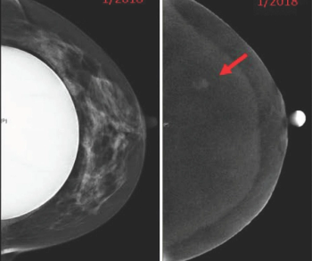
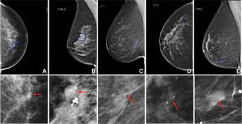
In a new study involving 198 contrast-enhanced mammography exams in 104 women with breast implants, researchers noted that only one patient had a complication.
In a multicenter study of 685 people who had positive findings for lung cancer on low-dose computed tomography (CT), researchers found that men, Black individuals, and smokers had lower rates of adherence with recommended follow-up care.
In two independent study cohorts, researchers found that first-degree relatives of probands with non-alcoholic fatty liver disease (NAFLD) and advanced fibrosis had a 14 to 15.6 percent higher risk of developing advanced fibrosis.

In a recent video interview, Thomas Marini, MD discussed the need for ultrasound access to facilitate breast cancer screening in underserved populations and the potential utility of volume sweep imaging, a handheld ultrasound technique that requires minimal training and has a high rate of agreement with conventional ultrasound on BI-RADS assessments.
Reviewing data from over 4 million patients who presented to emergency departments with suspected urinary stone disease between 2012 and 2018, researchers noted a greater than 10 percent decrease in visits with no imaging study and a greater than 10 percent increase in visits with CT imaging.
Emphasizing ease of installation and a cloud-based service, Cleerly said its new Proxy software allows smooth transmission of coronary computed tomography angiography (CCTA) scans between interdisciplinary clinicians caring for patients with heart disease.
Emerging research revealed that a deep learning model had a nearly twofold increase in successful segmentation and reconstruction of coronary total occlusions (CTOs) on coronary computed tomography angiogram (CCTA) and a 73 percent reduction in post-processing and measurement time in comparison to a conventional manual approach.
For patients undergoing stereotactic body radiotherapy (SBRT) for prostate cancer, the acute genitourinary (GU) toxicity rate associated with the procedure was 19 percent lower with magnetic resonance imaging (MRI) guidance in comparison to computed tomography (CT) guidance, according to new research presented recently at the American Society for Radiation Oncology (ASTRO) Annual Meeting.
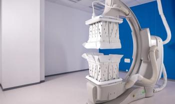
The automated head-to-toe shielding device, which reportedly blocks over 90 percent of radiation scatter to interventional radiologists and other clinicians, is now compatible with Siemens Artis fluoroscopy C-arm systems.
In separate test sets of Israeli women and United States women who had either ductal carcinoma in situ or invasive breast cancer, emerging artificial intelligence (AI) algorithms achieved an area under the curve (AOC) of 88 percent and 80 percent, respectively, for malignancy detection.
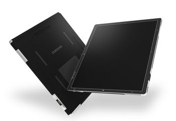
The AccE Glass-Free Detector reportedly combines high-resolution capability with a variety of benefits including improved functionality, enhanced visibility, and a lightweight design.
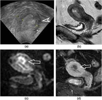
In a new study looking at pre-operative assessment of low-grade endometrial cancer, researchers found that while magnetic resonance imaging (MRI) had 20 percent higher specificity than transvaginal ultrasound for deep myometrial invasion, there was no difference in sensitivity.

In a recent video interview from the American Society for Radiation Oncology (ASTRO) Annual Meeting, Benjamin Lowentritt, MD discussed the challenges of conventional imaging in diagnosing prostate cancer recurrence and the potential of an emerging high affinity, radiohybrid prostate-specific membrane antigen/positron emission tomography (PSMA/PET) imaging agent.
Five weeks of radiation therapy is just as effective as eight weeks of radiation treatment for men with high-risk prostate cancer, according to new research presented at the American Society for Radiation Oncology (ASTRO) Annual Meeting.
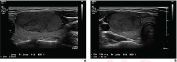
The retrospective study of patients 21 years of age or younger found that a deep learning algorithm and use of the American College of Radiology’s Thyroid Imaging Reporting and Data System (TI-RADS) both had more than a 26 percent greater sensitivity for differentiating thyroid nodules on ultrasound in comparison to radiologist assessment.
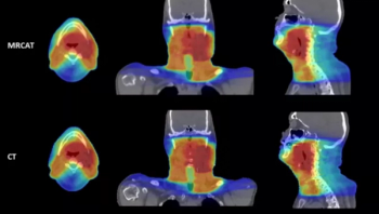
For physicians performing radiotherapy treatment of soft tissue tumors in the head and neck, the MRCAT Head and Neck offers an artificial intelligence (AI) application that allows the use of magnetic resonance imaging (MRI) as the primary or sole imaging for procedure planning.
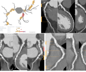
Plaque Analysis and RoadMap Analysis, two artificial intelligence (AI)-enabled assessment products, may enhance clinical evaluation of coronary artery disease (CAD) on cardiac computed tomography angiography (CCTA).
In a recent letter to U.S. Rep. Rosa DeLauro (D-CT), the Food and Drug Administration (FDA) said a final rule on amendments to the Mammography Quality Standards Act (MQSA), including an oft-delayed national standard for breast density notification in mammography reporting, may be published in the next couple of months.
The inclusion of digital bismuth germanate (BGO) detector material with the Omni Legend system reportedly more than doubles the sensitivity of older PET/CT devices, improves scan times, and enhances the detection of small lesions.
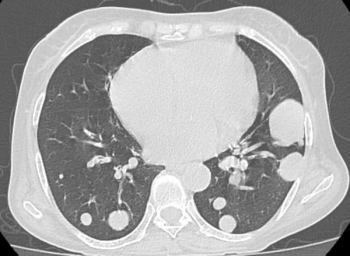
Out of 1,600 patients who had surgery for colorectal cancer, nearly 15 percent had pulmonary metastases within 15.4 months and higher-risk patients experienced lung metastases within three months, according to new research presented at the Scientific Forum of the American College of Surgeons Clinical Congress.
In a recent video interview, Andrew Trout, MD discussed key attributes and efficiencies in utilizing the Discovery MI Gen 2 digital positron emission tomography/computed tomography (PET/CT) system at the Cincinnati Children’s Hospital Medical Center.
A multi-year agreement with a large Chile-based supplier of raw iodine is part of GE Healthcare’s commitment to increase the production of iodinated contrast media, commonly used in computed tomography imaging, by 30 million annually in 2025.
An emerging radiomics model, derived from 68Ga-PSMA-11 PET, reportedly offers a superior area under the curve (AUC) and higher sensitivity than radiologist assessment in detecting intraprostatic lesions via positron emission tomography/computed tomography (PET/CT) in patients with prostate cancer.
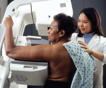
In a large retrospective review of over four million Medicare claims, researchers found that Black women were 16 percent less likely to have access to digital breast tomosynthesis than White women.
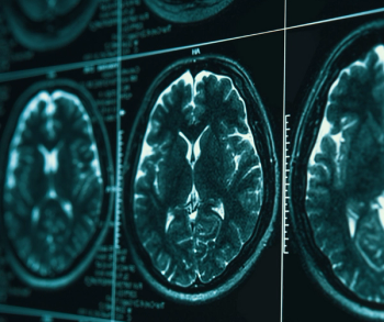
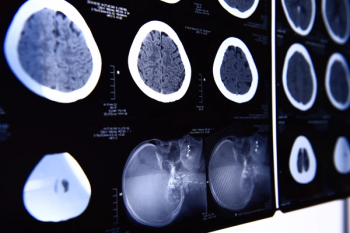
While current consensus guidelines do not recommend the use of brain MRI screening in patients with breast cancer, a new study shows significantly elevated risks for the development of central nervous system metastasis in patients with inflammatory breast cancer.
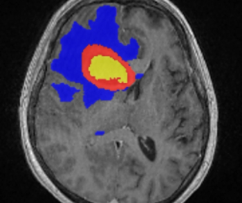
Incorporating artificial intelligence (AI)-based technology, Neosoma HGG reportedly demonstrated a 95.5 percent accuracy rate in measuring brain tumor volume on brain magnetic resonance imaging (MRI) scans at various points during the treatment of patients with high-grade gliomas.
In a wide-ranging video interview, Wendie Berg, MD, PhD, discussed her family history with breast cancer, the founding of DenseBreast-info.org, emerging research with contrast-enhanced mammography and the need for a national standard on breast density notifications.
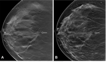
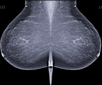
In a large study of nearly 100,000 women, researchers found that the combination of digital breast tomosynthesis (DBT) and synthesized mammography had more than triple the detection rate for invasive breast cancer in extremely dense breasts in comparison to digital mammography alone.

TeraRecon Neuro reportedly offers six automated and customizable computed tomography (CT) perfusion maps that facilitate assessment of brain function in hemorrhagic and ischemic neurological cases.