How Photon-Counting CT Helps Us Recognize that Coronary Stenosis is Just One Piece of the Puzzle
While a new article raises provocative questions about the capability of photon-counting CT (PCCT) to perhaps replace diagnostic ICA, the real revolution with this modality is going beyond stenosis measurement to a renewed focus on the disease of atherosclerosis.
For decades, coronary imaging has been dominated by one question: How severe is the stenosis?
However, coronary atherosclerosis is much more than luminal narrowing. A new article by Antoniades, Kotronias and Blankstein in the European Heart Journal explores how photon-counting computed tomography (PCCT) could fundamentally change cardiovascular imaging.1
Five Ways PCCT Is Enhancing Cardiac CT
I believe the implications go far beyond better image quality. Photon-counting CT is progressively expanding what we can investigate in a single examination.
• Coronary stenosis. Ultra-high spatial resolution improves luminal assessment, particularly in heavily calcified arteries and stents.
• Atherosclerotic plaque. There is more accurate morphological characterization and quantitative assessment of plaque burden.
• Coronary inflammation. Perivascular adipose tissue analysis offers information beyond anatomy with the potential for more sophisticated spectral characterization.
• Coronary physiology. CT-derived functional assessment can complement anatomical findings.
• Myocardial characterization. Extracellular volume mapping and late iodine enhancement open new opportunities to assess myocardial injury and tissue abnormalities.
Reshifting the Focus to Addressing the Disease of Atherosclerosis
The authors of the article raise two provocative questions:
Could PCCT eventually replace diagnostic invasive coronary angiography?
Could PCCT reduce the need for cardiac MRI in selected clinical scenarios?
These are legitimate questions, but I believe the real revolution is even more fundamental. For too long, we have approached coronary artery disease primarily as a problem of stenosis, but stenosis is not the disease.
Luminal narrowing is only one of its morphological consequences. Ischemia and myocardial injury are possible downstream consequences.
Photon-counting CT offers the opportunity to investigate several dimensions of this disease within the same examination.
Of course, technological capability is not equivalent to demonstrated clinical benefit. Quantitative biomarkers require validation, and better images do not automatically translate into better outcomes. However, the direction is clear.
The future of cardiac CT is not simply about competing with invasive angiography in measuring coronary stenoses. It is about moving from stenosis-centered imaging to disease-centered imaging. The ultimate goal should not be measuring a narrowing more precisely. It should be better understanding of the disease and making better clinical decisions.
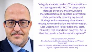
Dr. Cademartiri is the director of advanced cardiovascular imaging and photon-counting CT at the Scientific Institute for Research, Hospitalization, and Healthcare Synlab Diagnostic Network in Naples, Italy. He is also a consultant in advanced cardiovascular imaging at CDI/Centro Diagnostico Italiano in Milan, Italy.
Reference
- Antoniades C, Kotronias RA, Blankstein R. Photon-counting computed tomography: next-generation imaging of coronary stenosis, inflammation, and myocardial status. Eur Heart J. 2026 Sep 18:ehaf890. doi: 10.1093/eurheartj/ehaf890 .
(Editor’s note: This blog is adapted with permission from Dr. Cademartiri’s original LinkedIn post available at:
Related to this article