
Blog|Articles|August 21, 2026
Could Photon Counting CT Enhance Local Staging of Bladder Cancer?
Author(s)Filippo Cademartiri, MD, PhD
Reducing false positives in evaluation of muscle invasion in bladder cancer cases that can occur with conventional imaging, spectral information with photon-counting CT may offer significantly improved accuracy and specificity in local staging.
Advertisement
Conventional CT has long been considered limited for local staging of bladder cancer, particularly for distinguishing non–muscle-invasive from muscle-invasive disease. A new prospective study suggests that photon-counting CT may start changing that assumption.
In 110 patients with pathologically confirmed bladder cancer, the study authors compared low-keV virtual monoenergetic images with conventional-like polychromatic reconstructions. Among 40–70 keV datasets, 40-keV VMI provided the highest tumor signal-to-noise ratio (SNR) and contrast-to-noise ratio (CNR), and the best delineation of the tumor–bladder wall interface.1
More importantly, this translated into diagnostic performance. Across three readers, 40-keV VMI achieved:
• accuracy between 80.9 to 87.3 percent;
• sensitivity between 88.9 to 96.3 percent; and
• specificity between 78.3 to 86.7 percent.1
Accuracy and specificity were significantly higher than with conventional-like images while sensitivity remained similar. Diagnostic confidence also increased substantially.
What is the Critical Point of the PCCT Study?
This is not simply a study showing that low-keV images look better.
The important result is that spectral information changed diagnostic performance, particularly by reducing false-positive assessment of muscle invasion.
However, there is an important limitation. Even with PCCT, the individual bladder wall layers were not directly resolved. Improved staging resulted mainly from greater iodine-related tumor-to-wall contrast and better visualization of the lesion base and ancillary features, not direct visualization of the muscularis propria.
This study was also a single-center, single-vendor study with only 27 muscle-invasive cancers and no direct comparison with MRI.
However, from my perspective, this is exactly the type of PCCT study we need. This is not another demonstration of lower noise or prettier images. Instead, we see evidence that the technological advantages of photon counting can change a clinically relevant diagnostic decision.
The study authors appropriately do not propose replacing MRI.
Instead, the opportunity is much more pragmatic: extracting local staging information from a CT urography examination that the patient is already undergoing without additional radiation or contrast administration.
Where PCCT Enters the Picture
This study illustrates an important evolution of PCCT going from image quality improvement to clinical value creation.Higher spatial resolution plus spectral information plus low-keV imaging can potentially transform examinations we already perform into richer diagnostic tools.
That may ultimately be one of PCCT's greatest strengths. It’s not necessarily performing more imaging but extracting more clinically actionable information from the same examination.
Dr. Cademartiri is the director of advanced cardiovascular imaging and photon-counting CT at the Scientific Institute for Research, Hospitalization, and Healthcare Synlab Diagnostic Network in Naples, Italy. He is also a consultant in advanced cardiovascular imaging at CDI/Centro Diagnostico Italiano in Milan, Italy.
(Editor’s note: This blog is adapted with permission from Dr. Cademartiri’s original LinkedIn post at:
Reference
- Wang W, He J, Wu J, et al. Photon-counting CT with low-keV virtual monoenergetic images for detecting muscle invasion in bladder cancer: a prospective study. AJR Am J Roentgenol. 2026 Aug 19. doi: 10.2214/AJR.26.35310.
Advertisement
Related to this article

Catch up on the top radiology content of the past week.

While a new article raises provocative questions about the capability of photon-counting CT (PCCT) to perhaps replace diagnostic ICA, the real revolution with this modality is going beyond stenosis measurement to a renewed focus on the disease of atherosclerosis.
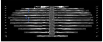
The use of adjunctive CT bone reading software in women with breast cancer detected between 8 to 22 percent more metastatic lesions for reviewing radiologists and reduced CT interpretation time by 50 percent or more, according to a new study.
Catch up on the top radiology content of the past week.

Magnetic resonance imaging offered a 29 percent higher sensitivity than CT for differentiating liver metastases, but CT provided 12 percent higher sensitivity than MRI for assessing vascular invasion, according to a new 40-study meta-analysis.

Does fractional flow reserve (FFR) or quantitative plaque burden from CCTA provide greater prognostic insight into coronary disease?

Catch up on the most-well viewed radiology content in September 2026.
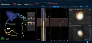
The FFR-IQ software recently demonstrated a 92.2 percent sensitivity for non-invasive estimation of fractional flow reserve (FFRC) based on coronary CT angiography (CCTA) scans.
Advertisement
Advertisement
Trending on Diagnostic Imaging
1
FDA Clears AI-Enabled FFR-IQ Software for Estimating Fractional Flow Reserve from CCTA
2
FDA Clears MRI-Based AI Software for Prognostic Evaluation of Patients with Mild Cognitive Impairment
3
Can the Kaiser Score with Breast MRI Improve Clarity for BI-RADS 0 and 4 Assessments on Mammography and Ultrasound?
4
The Reading Room Podcast: Emerging Concepts with AI Integration in Radiology
5