


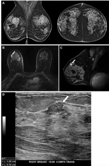
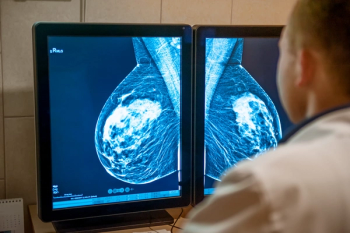
New research suggests the capability of a mammography-based deep learning model to identify women at high risk of breast cancer led to more than triple the cancer detection rate on breast MRI in comparison to traditional risk assessment tools.
New research suggests the capability of a mammography-based deep learning model to identify women at high risk of breast cancer led to more than triple the cancer detection rate on breast MRI in comparison to traditional risk assessment tools.
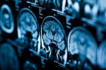
The artificial intelligence (AI)-enabled AIRAscore can reportedly provide quantitative brain volume data within five minutes of assessing brain MRI scans.
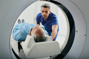
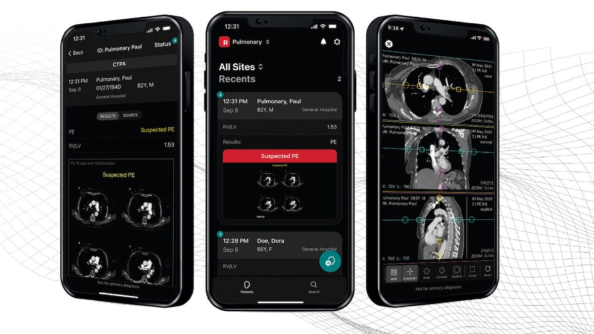
The use of automated detection of large vessel occlusion on computed tomography (CT) through artificial intelligence (AI) software reportedly led to an 11.2-minute reduction in triage time from the completion of imaging to initiation of endovascular therapy, according to newly published research.

Catch up on the top radiology content of the past week.
Indicated for the triage and notification of obstructive hydrocephalus on non-contrast brain computed tomography (CT), the artificial intelligence (AI)-enabled software is reportedly the first radiology triage modality to obtain the Food and Drug Administration’s (FDA) Breakthrough Device Designation.
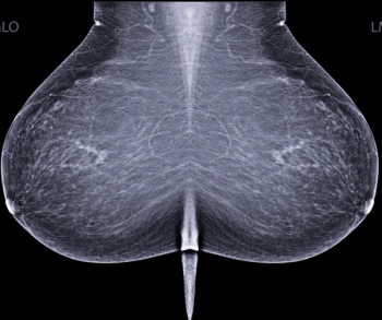
In a prospective study of over 55,000 women who had screening mammography, researchers found that double-reading by a radiologist and artificial intelligence (AI) was non-inferior to double-reading by two radiologists in detecting breast cancer.
Catch up on the top radiology content of the past week.
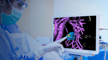
Through the use of artificial intelligence (AI) and imaging modalities such as ultrasound, CT, and MRI, the newly FDA-cleared VisAble.IO software reportedly enhances planning and real-time assessment for liver tumor ablation procedures.
In separate test sets that included challenging mammography cases, researchers found that artificial intelligence (AI) demonstrated similar sensitivity and specificity for detecting breast cancer in comparison to assessments from over 500 clinicians.
Catch up on the top radiology content of the past week.
In the third episode of a three-part podcast, Anand Narayan, M.D., Ph.D., and Amy Patel, M.D., discuss the challenges of expanded breast cancer screening amid a backdrop of radiologist shortages and ever-increasing volume on radiology worklists.
Catch up on the top five most viewed content at Diagnostic Imaging in August 2023.
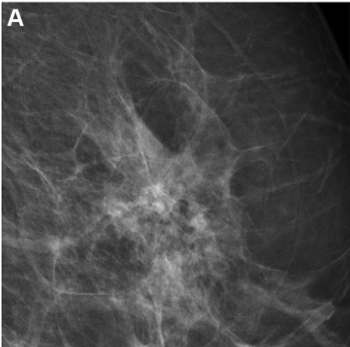
The study demonstrated that the combined model yielded improved risk assessment for both interval and long-term breast cancers.
Catch up on the top AI-related news and research from the past month.
Catch up on the top radiology content of the past week.
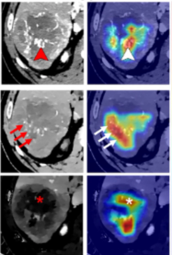
In comparison to clinical-radiologic assessment, a deep learning CT radiomics nomogram had a 10 percent higher AUC and a 27 percent higher specificity for predicting the macrotrabecular massive subtype of hepatocellular carcinoma in external data testing.
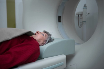
The Echelon Synergy MRI system reportedly uses deep learning technology to accelerate image acquisition and enhance image quality.
Catch up on the top radiology content of the past week.
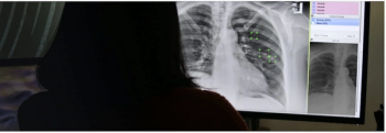
The artificial intelligence (AI)-enabled VisiRad XR reportedly demonstrated an 83 percent sensitivity rate for detecting lung nodules and masses on chest X-rays in one retrospective study.
The De Novo approval for Viz HCM, which assesses electrocardiograms with artificial intelligence (AI) to identify possible cases of hypertrophic cardiomyopathy (HCM), is the 12th FDA clearance of algorithms on the Viz.ai Platform.
Catch up on the top radiology content of the past week.
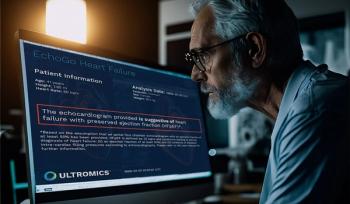
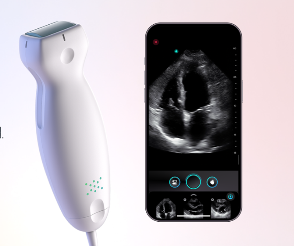
In what may represent the first CMS approval of a new technology add-on payment (NTAP) for an artificial intelligence (AI)-based heart failure detection platform, use of the EchoGo Heart Failure system will be eligible for up to $1,023.75 in NTAP reimbursement per acute hospital in-patient stay as of October 1, 2023.
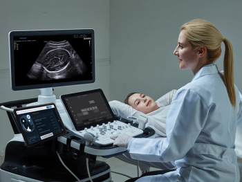
With reported performance validation on over 17,000 ultrasound images, Sonio Detect employs artificial intelligence (AI) to help ensure quality criteria for fetal ultrasound imaging of the brain and heart.
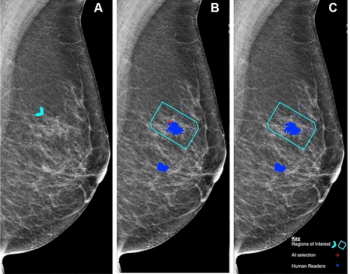
Reportedly the first randomized trial to examine the impact of artificial intelligence (AI) on screening mammography, researchers found AI-aided screening led to a 20 percent increase in breast cancer detection and a 44.3 percent decrease in mammography screening workload.

Catch up on the top AI-related news and research from the past month.