


A five-tiered approach to cross-sectional interventional procedures can help radiologists determine which patients to treat first, minimizing likelihood of viral transmission.
A five-tiered approach to cross-sectional interventional procedures can help radiologists determine which patients to treat first, minimizing likelihood of viral transmission.
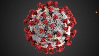
Researchers from Mount Sinai examine and assess the imaging modalities used to evaluate patients positive for the virus.

Diagnostic Imaging's Weekly Scan: May 29, 2020
Research shows 77 percent of children who are confirmed positive for the virus have no findings via chest CT.
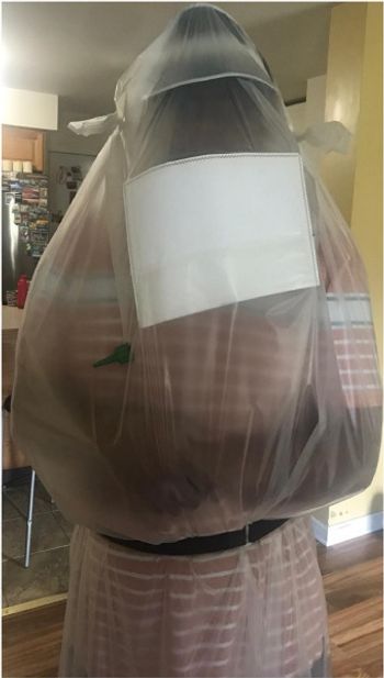
Patient isolation bag could not only reduce risk of viral transmission, but could also streamline CT workflow.
Radiology department staff have the ability – and responsibility – to calm patient anxieties about imaging studies and the virus during the pandemic, says one radiology supervisor.

Software supports CT and X-ray chest imaging.
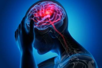
Half of patients hospitalized who have neurological findings for acute stroke could die.
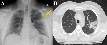
Research reveals lung findings not typically associated with viral pneumonia.
Model could help detect the virus, isolate patients, and prevent disease spread.
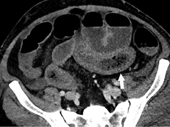
Results show COVID-19-positive patients with BMI greater than 30 are at significantly higher PE risk.

Diagnostic Imaging's Weekly Scan: May 15, 2020
Radiologists must continue to be aware that patients obtaining scans for non-respiratory symptoms could still present findings of COVID-19.
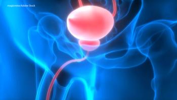
Researchers identify 18F-PMSA-1007 as an equally effective radiopharmaceutical for detecting prostate cancer.
Algorithms can create errors in multiple imaging systems, according to new tests.
Affected patients were sicker and more likely to be admitted to the ICU.
Concentration on virus-related scans largely sidelines other imaging needs.

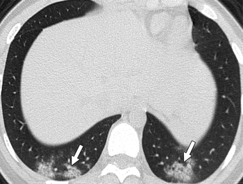
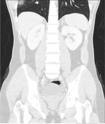
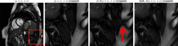
Tool pinpoints incidental findings on CT scans that contain the lung or part of the lung.
Emergency medicine expert panel outlines support for using point-of-care ultrasound with patients with suspected infection.
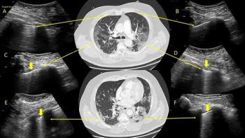
Side-by-side images show lung ultrasound pinpoints same findings as low-dose chest CT.
COVID-19 and other viral infections have unique imaging characteristics that can help with diagnosis, experts say.

Diagnostic Imaging Weekly Scan -- May 1, 2020
System uses ultraviolet light system to kills advanced viruses and bacteria in minutes.
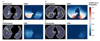
Adding AI to manual reads improves accuracy, specificity, and sensitivity.
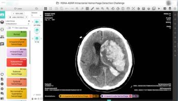
Together, RSNA and the American Society of Neuroradiology launched the largest collection of expert-annotated brain hemorrhage CT scans.