


New case volume-based training curriculum is designed to create a standardized competency level for radiologists-in-training.
New case volume-based training curriculum is designed to create a standardized competency level for radiologists-in-training.

CT can provide a diagnosis in a less invasive way and without a high false-positive rate.
Cerebral Aneurysms and CT Angiography; MammoScreen and Breast Cancer Detection; Low-Dose Lung Cancer Screening Program Performance; and Breast Cancer Screening Advocacy Efforts
In a study from China, there was no statistical significance in cancer detection rates between high-risk patients who were screened and those who were not.

Purchasing vouchers for imaging services could present safety risks and set customer up for upselling attempts.

Deep learning tool improves cerebral aneurysm detection, specifically among radiologists with fewer years’ experience.
In this podcast episode, Dr. Shalom Kalnicki, from Montefiore and Albert Einstein College of Medicine, discusses the disparities minority patients face with cancer screenings and what can be done to increase access during the pandemic.
Ali Gholamrezanezhad, M.D., from the Keck School of Medicine, shares his COVID-19 insights after conducting more than three dozen studies.

Breast Cancer Screening in Indian & Pakistani Women; Fluciclovine PET for Prostate Cancer Imaging; Cardiac Ultrasound & COVID-19; and Improving Mammography Patient Experience

An analysis of the Dallas Heart Study from UTSW details how use of CAC scoring can help determine what patients would stand to benefit from aspirin use for primary prevention.
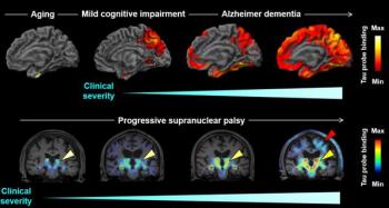
Modifying an existing imaging probe and labeling it with fluorine-18 improves providers’ ability to pinpoint protein accumulation – and differentiate between neurodegenerative conditions.

The scan, which is already part of the stroke management process, offers an opportunity for faster identification of patients with viral infection.
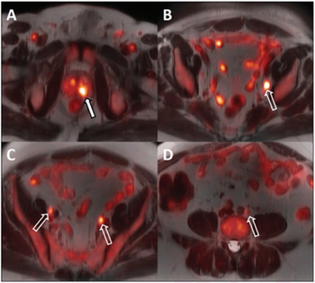
18F-fluciclovine PET/MRI can improve treatment guidance with better staging and evaluation of androgen deprivation therapy.

Ultrasound as the New Stethoscope; Pre-Operative MRI and Dense Breasts; Spectral CT and COVID-19; Corporatization in Radiology
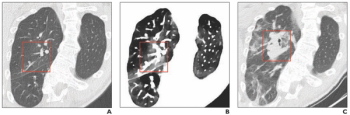
Spectral CT provides greater detail about the extent of ground-glass opacities than conventional CT scans.
African American, as well as other racial and ethnic minority, patients likely have lower levels of CTC screening due to out-of-pocket costs.
Training a neural network with images captured by dual-energy CT can produce high quality studies without the added dose or expense.
The strategy could be a less expensive alternative to CT when evaluating pulmonary fibrosis and pulmonary edema.
Opposition to AI in Mammography; Alzheimer's Disease and Vascular Dysfunction; Clear & Present Danger for The Match; Trends & Innovations in Breast Imaging

In this podcast, Dr. Anupam Basu from Cook County Health in Chicago discusses the 30-pack-year threshold for lung cancer screening that overlooks at-risk African American smokers.
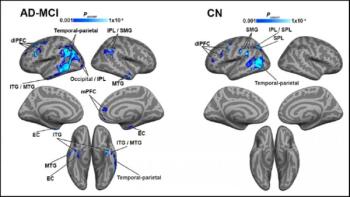
As Alzheimer’s disease progresses, more brain regions exhibit evidence of this link.

Collaborative Imaging’s Dhruv Chopra, MBA, takes a deeper look into how many follow-up recommendations are ignored and what can be done to reverse the trend, leading to better patient care.
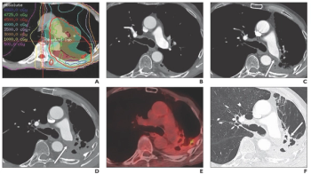
Pulmonary artery thrombosis presents differently from acute pulmonary embolism on CT scans.

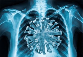
SARS-CoV-2 virus found inside CT scanner used on high volume of patients with viral infection.

Dual-Energy CT's Impact in the Emergency Department; Mass Casualty Planning; Language Barriers to Mammography for Spanish-Only Speakers; Improving the Patient Experience for Mammography During the Pandemic