
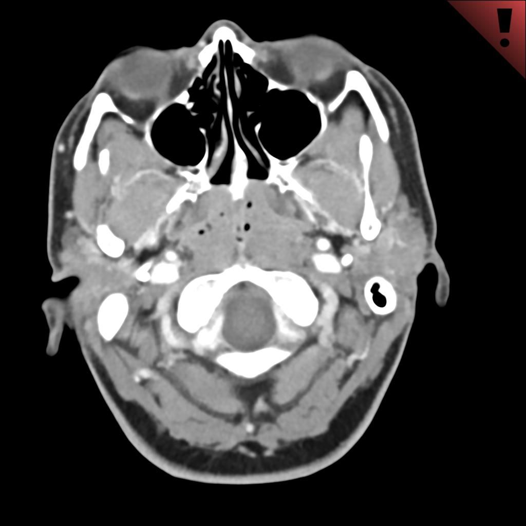














Case History: 45-year-old patient presented with complaints of vomiting for a few days.

Case History: 45-year-old patient presented with complaints of vomiting for a few days.

Case History: 43-year-old female with no significant medical or surgical history presents with acute onset, severe left lower abdominal pain.

Computed tomographic imaging of the colon demonstrates findings predictive for recurrent diverticulitis.

Case History: 40-year-old patient presented with history of fever and hemoptysis.

Case History: 57-year-old patient with longstanding history of swelling on left side of face and difficulty opening mouth.

CT combined with 3D printing accurately reproduced damaged parts of the middle ear.

CT shows former NFL players may have enlarged aortas compared with controls.
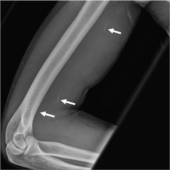
Radiologists in the emergency department can help advocate for patients with opioid-use disorders.

Case History: 45-year-old patient with laryngeal cancer presented with history of breathlessness, hemoptysis, increased creatinine.

Abdominal CT studies can help clinicians accurately evaluate the lumbar spine.
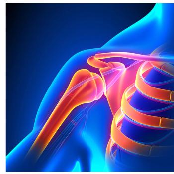
Use of imaging for musculoskeletal extremities has increased significantly over the past 20 years.
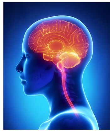
Older patients with brain PET results showing elevated amyloid have many questions.

Case History: 55-year-old with pain and swelling in left submandibular region.

The PanCan approach to lung cancer detection utility in lung cancer screening.

Effect of breath holding during lung PET/CT.

Nuclear medicine radiologic technologists may be at risk for developing cataracts.
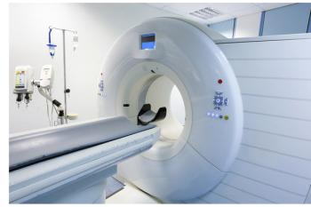
Multiple CT scans for young adults with non-traumatic acute abdominal conditions increase radiation exposure.

More patients and providers are aware of CT dose in 2015 than they were in 2004.
Disparities in radiation doses from CT scans in different countries.
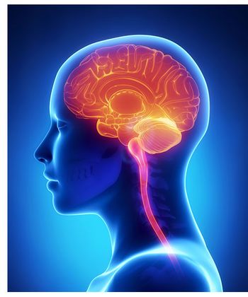
FDG imaging vs PET/MR for diagnosing children with suspected localization-related epilepsy.

Case History: 13-year-old male with complaint of headache and hearing loss.

Case History: 40-year-old female presented with history of epigastric fullness associated with epigastric and left flank pain and burning micturition.
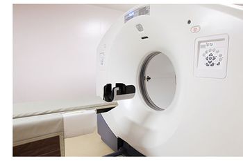
Using PET/CT imaging to evaluate patients following chemoradiotherapy for anal cancer can alter patient management choices.
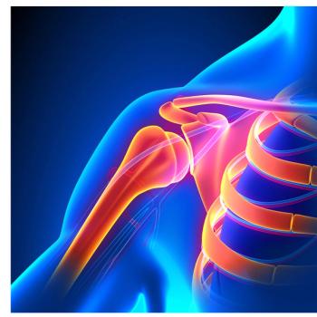
Lower doses of radiation during CT scans for extremities still provide good image quality.

Cranial CT scans performed on children in non-pediatric hospitals expose them to more radiation.