


Point-of-Care Ultrasound and COVID-19, Plus MRI and Schizophrenia, Pediatric Cancer, and Alzheimer's
Diagnostic Imaging's Weekly Scan: July 3, 2020

Diagnostic Imaging's Weekly Scan: July 3, 2020
Monitoring color flow gain in 3D quantitative blood flow measurements provides accurate and reliable information.

Database intended to be the largest open database of COVID-19-related imaging worldwide.

Amazon Alexa skill RAD-Assistant successfully retrieves pulmonary nodule and ovarian cyst clinical follow-up guidelines.
Multisystem Inflammatory Syndrome in Children appearing in pediatric patients with COVID-19 infection and exposure.
Combining sound waves and a novel drug can reduce cancer cells by nearly 50 percent.
One rural-based model shows bringing interventional radiology services to older patients in their home not only improve access to care, but also catapults patient satisfaction.

AI model can help alleviate increased sonographer workload, but still has high missed diagnosis rate.

Diagnostic Imaging's Weekly Scan: June 5, 2020
The modality is an effective, reliable alternative to chest CT or chest X-ray in this vulnerable population.

A five-tiered approach to cross-sectional interventional procedures can help radiologists determine which patients to treat first, minimizing likelihood of viral transmission.
Researchers from Mount Sinai examine and assess the imaging modalities used to evaluate patients positive for the virus.
Diagnostic Imaging's Weekly Scan: May 22, 2020

Research reveals lung findings not typically associated with viral pneumonia.

Diagnostic Imaging's Weekly Scan: May 15, 2020

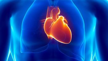
Clearance allows for applications for lung and cardiac complications.
Adhering to a policy of using lung ultrasound as an initial scan reduced the use of other imaging options.
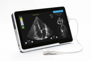
AI-guided imaging supports assessment of cardiac function and reduces personnel COVID-19 exposure.
Affected patients were sicker and more likely to be admitted to the ICU.
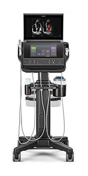
Philips releases new transducer with 30-percent greater penetration for more-detailed images.

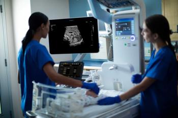
Emergency medicine expert panel outlines support for using point-of-care ultrasound with patients with suspected infection.
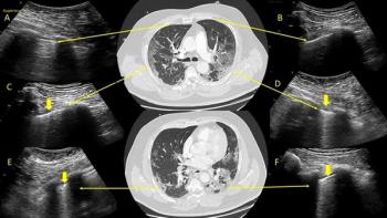
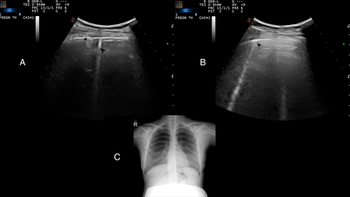
Side-by-side images show lung ultrasound pinpoints same findings as low-dose chest CT.
After 2.5 hours, providers could successfully identify lungs with pathological patterns indicative of viral infection.

Agency outlines modifications that will and won’t be accepted without approval during the outbreak.
Reduced radiation exposure and virus transmission is possible for both patients and providers.