


Helping ultrasound meet its potential.
Helping ultrasound meet its potential.

Adding automated breast US to regular screening of women with BRCA mutation and cancer detection.
Case History: 55-year-old with pain and swelling in left submandibular region.

FDA approves Philips’ ‘Small Parts’ ultrasound transducer.
Utility of both PI-RADS version 2 and MRI-ultrasound fusion biopsy for diagnosing prostate cancer.
Case History: 24-year-old male with enlarged cervical lymph nodes associated with weight loss and fever.
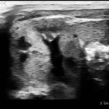
Case History: 49-year-old female presented with palpable right breast retroareolar mass.

The first choice for imaging adult patients with suspected appendicitis varies according to patient demographics and resource availability.

Ultrasound as an artform.

Using ultrasound as part of the FAST assessment for children with blunt abdominal trauma does not appear to improve quality of care.
Subsequent ultrasounds are more likely to occur when non-radiologists read initial ultrasounds in the emergency department than when radiologists read.
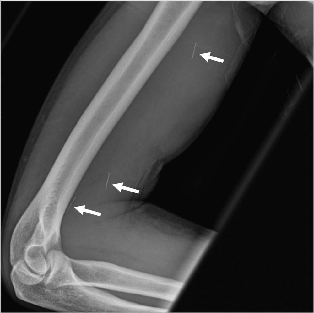
Radiographs can provide anatomic evaluation and appropriate starting point when investigating possible musculoskeletal infection.
Researchers determine quantitative ultrasound could be superior to conventional ultrasound for predicting fat acculturation in the liver.
Breast images performed outside cancer centers may be interpreted differently if reinterpreted at a cancer center.
Clinicians can use ultrasound to detect pediatric appendicitis, but not if the organ has perforated.

There is a higher chance that a follow-up examination will follow when nonradiologists interpret ultrasounds in the ER.
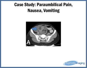
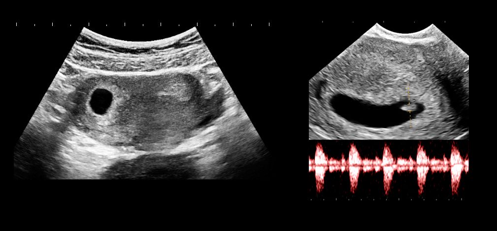
Case History: 35-year-old female presented with pain in the left paraumbilical region for two days, nausea and vomiting.
Appendiceal sonograms may help clinicians tell which patients have uncomplicated versus complicated appendicitis.

Rate of diagnoses and cost following thyroid imaging after benign fine-needle aspiration.
Trends in ultrasound from RSNA.
Ultrasounds repeated because of incomplete visualization of the fetus and detection of fetal abnormalities.

RSNA 2016 displays more signs pointing towards the blending of diagnostic, interventional, and therapeutic imaging.
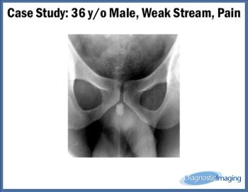
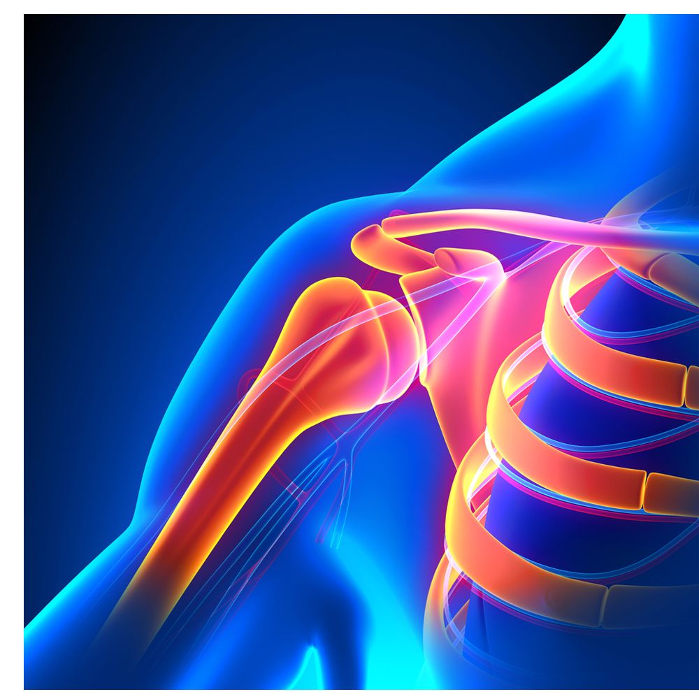
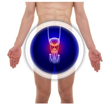
Case History: 36-year-old male presented with weak stream and pain.

How does accuracy compare in automated breast volume ultrasound and hand-held ultrasound?