














CT not always necessary for adult patients presenting with blunt trauma to the chest.
CT not always necessary for adult patients presenting with blunt trauma to the chest.

When CT is available in the emergency room, physicians are more confident about their patient care decisions.
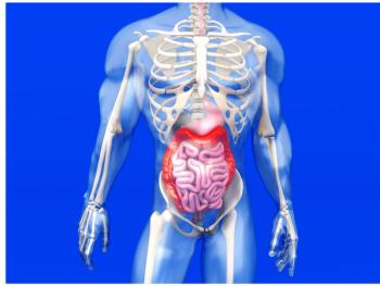
Using CT colonography for screening of colorectal cancer among Medicare patients is cost-effective.

Radiologists reading CT colonography may see difficult-to-detect polyps but not consciously register them.

Barco’s Coronis Uniti display system received expanded clearance for multi-modality viewing.
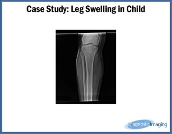
Case History: Pediatric patient presented with mild swelling of right lower leg.
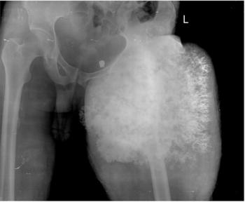
55-year-old male with swelling in left leg for four months.

CT performed on admission for patients with suspected acute appendicitis can predict hospital resource utilization.
Radiologists and nonradiologists must come to a consensus regarding who educates patients about radiation risks from diagnostic imaging.

Draft recommendations from the USPSTF on colorectal cancer screening omit CT colonography as a preferred screening method.
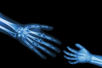
Use of ultrasound and MRI among children is rising, while CT imaging drops.
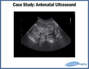
Case History: 25-year-old patient presents for antenatal ultrasound.

CCTA, when combined with calcium scoring, can rule out coronary artery disease in women with atypical chest pain.
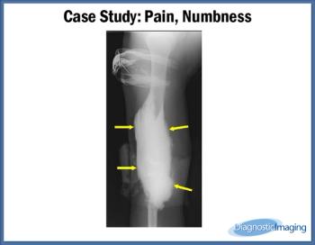
Case History: 57-year-old male with increasing pain and numbness following CT.

They might not be on the field, but radiologists are an important part of the NFL’s medical team.
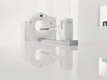
Brain images from FDG-PET can help detect Alzheimer’s disease in patients presenting with focal onset dementias.

Diagnostic imaging procedures are sometimes necessary for pregnant women, and can be done safely in some cases.
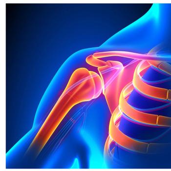
CT colonography to screen for colorectal cancer also gives physicians the opportunity to assess patients for osteoporosis.
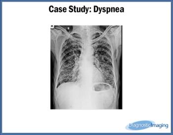
Case History: 74-year-old male smoking 48 packs/year with history of severe dyspnea.
Computed tomography angiography may show significant incidental findings that ED physicians should act upon.
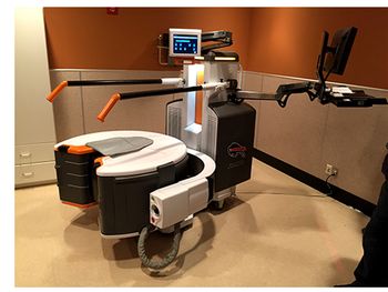
At AHRA 2015, Carestream showcased their investigational cone beam CT, but they’re not selling it to only radiologists.

CT scans for children with head injuries from sports should not be routine practice.
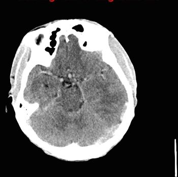
33-year-old male with history of cyanotic congenital heart disease status post multiple palliative cardiac surgeries.

This Q&A series explores radiology’s role in overdiagnosis in a variety of conditions. Here, we discuss mild traumatic brain injury.
FDG PET/CT scanning before treatment for invasive ductal carcinoma of the breast can predict survival.