


Access to imaging, particularly MRI and some forms of CT, vary considerably across the United States.
Access to imaging, particularly MRI and some forms of CT, vary considerably across the United States.
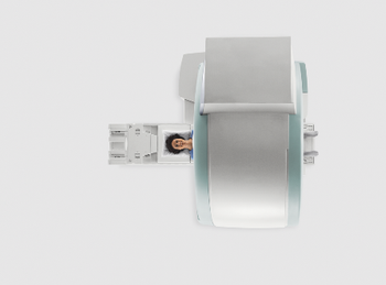
Patients undergoing MRI may have lessened anxiety if the procedure is well-explained.
MRI technique helps demonstrate the amount of damage in the gray matter of the brain is related to the severity of MS disability.
The reason for performing a breast MRI should be taken into consideration when assessing performance measures.
Patients are not given enough information about their implants and MRI safety.

A 72 year-old woman presents for routine screening mammogram with no complaints.
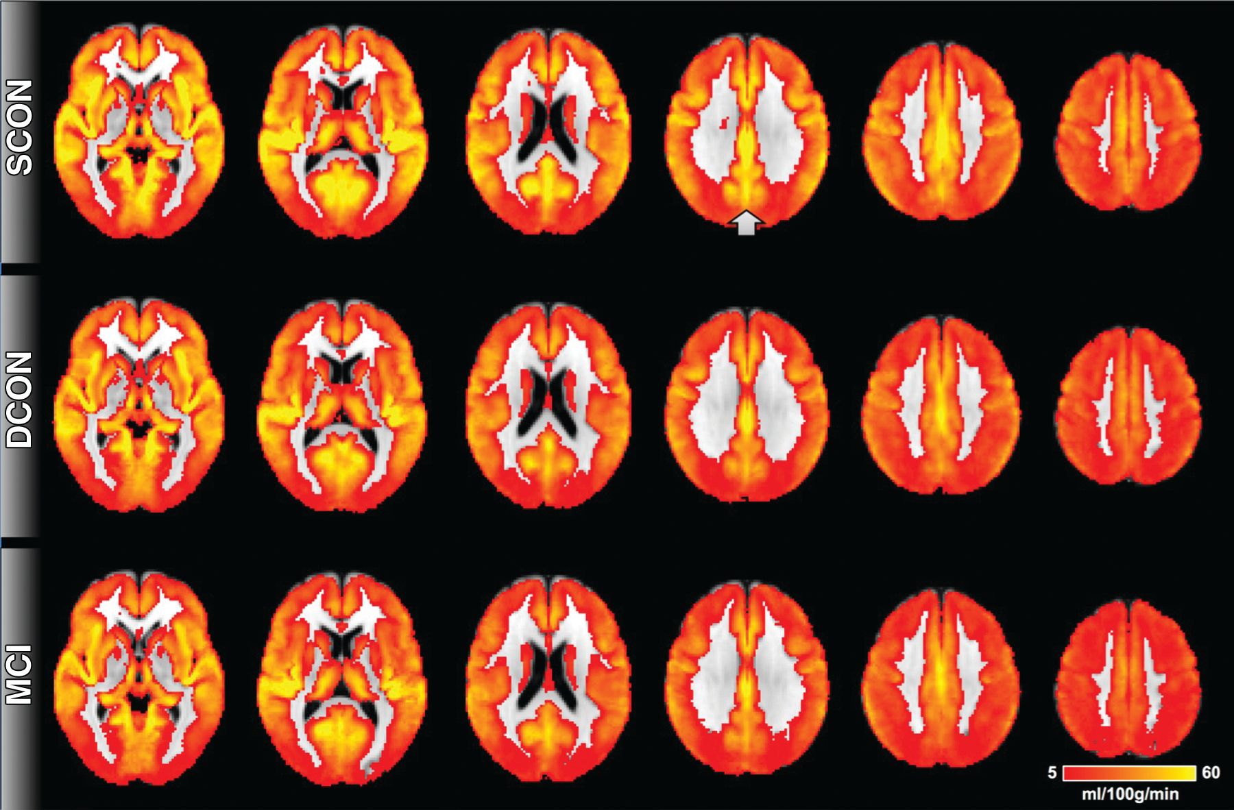
Magnetic resonance imaging on patients with multiple sclerosis who use a Will balance board showed changes of the brain that affected balance and movement.

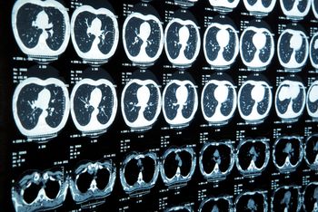
Emerging technology gives radiologists an unprecedented view of the brain.
Magnetic resonance imaging detected additional cancers among women who underwent breast conservation therapy following early diagnosis of breast cancer.
Multiparametric MRI helps identify low- and high-grade brain gliomas, reducing risk of inappropriate or delayed surgery.
Magnetic resonance imaging can be used to track brain growth of premature babies.
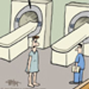
Difficult patients can get special treatment.
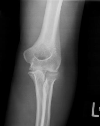
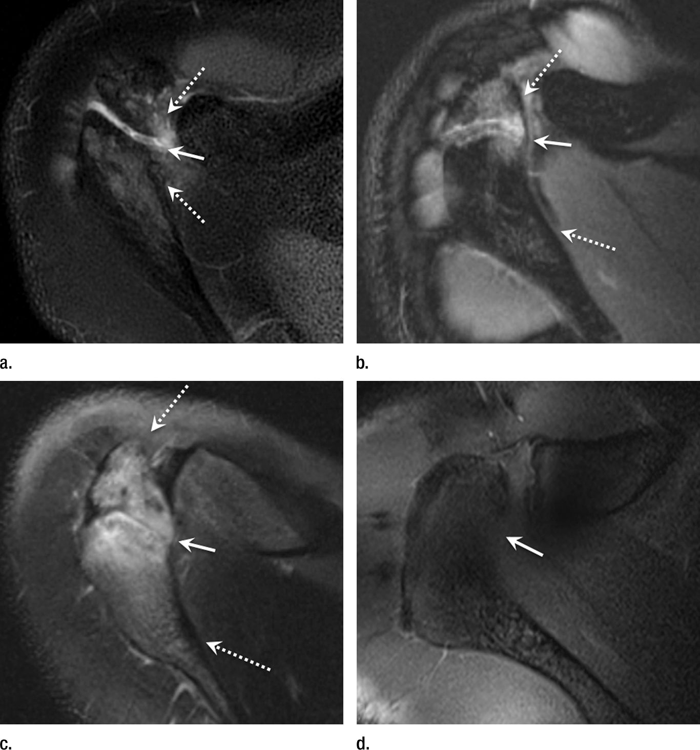
23-year-old male with a known undisclosed disease presents with left elbow pain.
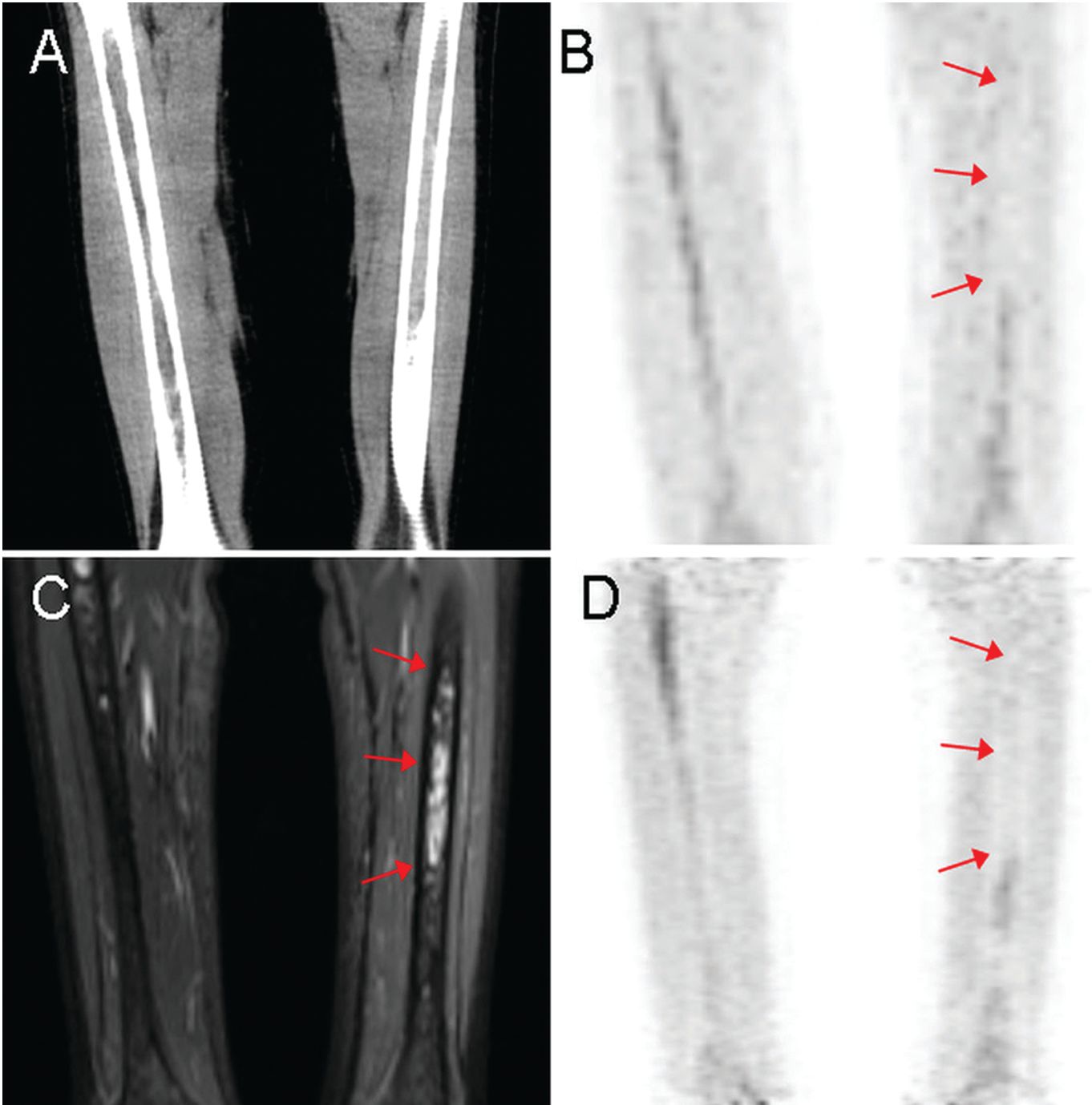
Subclinical inflammation of non-swollen joints not seen on radiographs can be detected on MRI.
Liver MRI is not accurate enough to definitely identify patients with iron overload.
Knee and shoulder MRI before radiography adds to patient care costs.
Use of imaging studies in emergency departments rose significantly from 1993 to 2007, but then declined from 2007 to 2012.

Long-time proponent of ultrasound for breast screening, Kevin M. Kelly, MD, discusses what radiologists need to know.
General practitioners in Australia are increasingly ordering imaging tests, with an increase of 45 percent over the past decade.
As growth of imaging utilization slows, radiologists worry about the effect on reimbursement.
Magnetic resonance imaging of pregnant patients with suspected appendicitis improves resource use, study finds.
Magnetic resonance images reveal characteristics of breast cancer can differ depending on age.
Magnetic resonance imaging of brains of adolescents before and after the Boston marathon attack provides insight on risk factors for PTSD, study finds.
Use of MRI to visualize white matter may help clinicians predict progression of MS.
Study tackles why women with most treatable form of breast cancer pursue most aggressive prophylactic measures; MRI false-positives have no effect.