







Brain MRI could expand the number of stroke patients eligible for a potentially life-saving treatment, according to a new study, published online and in the December issue of Radiology.
Brain MRI could expand the number of stroke patients eligible for a potentially life-saving treatment, according to a new study, published online and in the December issue of Radiology.
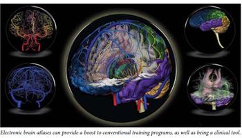
The topic of electronic brain atlases was first discussed in Diagnostic Imaging Asia Pacific almost a decade ago (see “Electronic brain atlases show value in brain studies,” June 2001, page 35). The article featured four atlases and addressed the potential of this innovation.
Every so often you come across an idea that has so much merit you can’t imagine why somebody hadn’t come up with it before. Such is the case with hospital gowns matched to a patient’s skin color. Although sky blue and forest green might be stunning on the right patient, it is hard to imagine either one helping docs spot signs of diseases that can be found in skin color changes.
A growing body of data is linking sportsrelated concussions to brain damage associated with cognitive impairment.
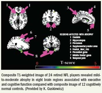
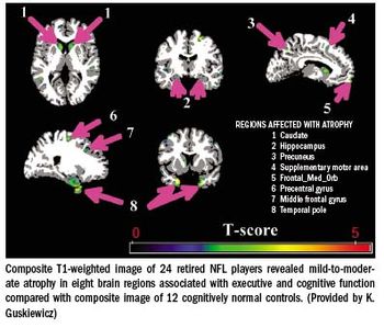
The brains of 24 retired U.S. National Football League players with known cognitive impairment show signs of damaging atrophy, according to advanced MRI studies described at the 2009 RSNA annual meeting.
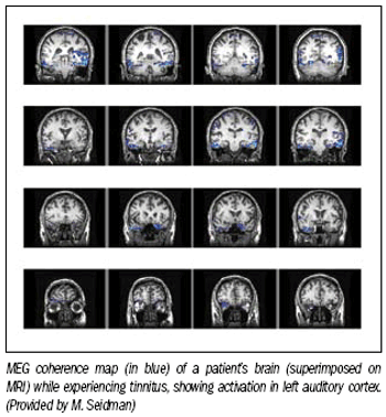
Researchers in Michigan are using magnetoencephalography of the brain to identify the source of tinnitus, a chronic ringing or buzzing in the ears or inside one's head.
Thanks to a near-perfect performance in a large cohort study in China, 64-slice CT angiography has shown it could replace digital subtraction angiography as the modality of choice for detecting suspected brain aneurysms at risk of spontaneous subarachnoid hemorrhage.
Thanks to a near-perfect performance in a large cohort study in China, 64-slice CT angiography has shown it could replace digital subtraction angiography as the modality of choice for detecting suspected brain aneurysms at risk of spontaneous subarachnoid hemorrhage.
Sometimes it’s just better not to know. One of those times may be when you feel perfectly fine, but your brain scan comes back with something that looks bad. The problem: how do you know it’s nothing to worry about?
Brain injuries invisible on CT and MRI, and apparent only in cognitive testing, can be revealed with diffusion tensor imaging, a study concludes.
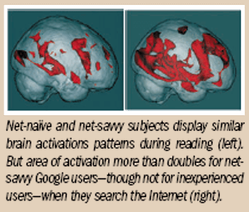
Net-naïve and net-savvy subjects display similar brain activations patterns during reading
Do you google? Relying on the Internet's most popular search engine has become second nature for frequent users.
An incidental finding of multiple sclerosistypelesions during brain MRI is no fluke. Anew study has found that some patientsdevelop the physical symptoms of the diseasewithin five years of the abnormalities'discovery on MRI.
An incidental finding of multiple sclerosis-type lesions during brain MRI is no fluke. A new study has found that some patients develop the physical symptoms of the disease within five years of the abnormalities’ discovery on MRI.
University of California, San Diego scientists are developing a new imaging modality that will study the body/brain dynamics of humans engaged in normal activity.
For victims of psychological trauma, the challenge of suppressing painful memories may be rooted in the wiring of the brain, an ongoing study revealed at the RSNA conference Wednesday.
Women with a weakened brain "reward circuitry" are at increased risk of weight gain over time and potential obesity, according to two studies from researchers at the University of Oregon. The risk increases for women who also have a gene associated with compromised dopamine signaling in the brain.
In a potential breakthrough study, French researchers used SPECT to identify brain abnormalities that present physiological evidence of fibromyalgia. The results quash the idea that diffuse, sometimes debilitating pain from the condition stems from anxiety and depression.
Cerebral blood flow imaging may eventually help identify young people who need preventive therapy Adolescents at relatively high risk for depression and alcohol abuse demonstrate distinct patterns of resting cerebral blood flow in areas of the brain associated with emotional behavior and decision making, according to preliminary results from the Research Imaging Center at the University of Texas Health Science Center in San Antonio.
Results from a decade-long study at the National Brain Aneurysm Center in St. Paul, MN, indicate that angiography to monitor intracranial brain aneurysm clip placement is safe and has altered management in more than one of 10 cases.
Functional diffusion technique shortens time needed to determine treatment efficiency, make adjustments The first three months after standard radiation therapy for a brain tumor must be hell for patients and their families. The established MacDonald criteria for assessing treatment force them to wait up to 10 weeks for follow-up CT or MR to determine whether the treatment is working.
Assuming you can do the math, your brain may operate more efficiently if you have first taken Ritalin (methylphenidate), a drug intended to help patients with attention-deficit hyperactivity disorder.
A new study from the University of Michigan featuring proton MR spectroscopy has found a key linkage between the widespread muscle pain, tenderness, and fatigue associated with fibromyalgia and brain glutamate. Findings could lead to new drugs to treat the condition, researchers said.
To the listener, jazz improvisation is an aural flight of fancy, borne aloft by a musician's on-the-spot skill and imagination. But functional MRI results show the brain actually follows a grounded process of activation and deactivation during these spontaneous musical riffs, according to researchers from the National Institutes of Health and Johns Hopkins University.
Neuroimaging with MRI at 3T is superior for nearly every application in the brain and spine, and it is certainly inferior for none. The technique has unique strengths for performing vascular work and functional brain imaging, but there is nothing that a 3T MR scanner can't do better than a 1.5T machine.