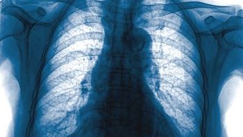
Using a deep learning-based automatic detection algorithm can identify more lung cancers on chest X-ray, potentially avoiding unneeded chest CT and accelerating treatment.
Whitney J. Palmer has been with Diagnostic Imaging since 2011, serving as the Senior Editor since November 2019. She has 20 years experience in healthcare and academic medicine reporting.
Using a deep learning-based automatic detection algorithm can identify more lung cancers on chest X-ray, potentially avoiding unneeded chest CT and accelerating treatment.

What department and practices leaders should focus on to be most successful post-COVID
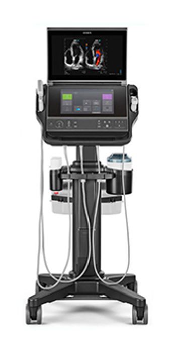
Sonosite PX improve image quality and workflow at the patient’s bedside.

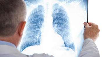
Concentrating on current smokers and those without positive CT results can improve patient participation and cost effectiveness.

Voice recognition artificial intelligence tools are taking on a growing role in radiology.
The pandemic is having several short- and long-term impacts that could change the face of private radiology practice.

Bedside lung ultrasound can be used to pinpoint which emergency department patients are more likely to die from viral infection.
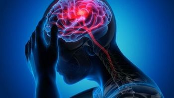
Avicenna.AI’s CINA Head earns 510(k) clearance for emergency room triage of intracranial hemorrhages and large vessel occlusions.
From virus behavior, to patient impact, to imaging implementation – what we know so far.
Maximizing Your COVID-19 Recovery, Re-Vamping the Radiology Residency Interview, Cardiac CT's Double Role with Osteoporosis, and the Human Impact of AI.
University Hospitals Cleveland Medical Center/Case Western Reserve saw a 55-percent overall imaging volume drop during the pandemic and unearthed some commonalities with other, harder-hit areas.
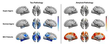
Images reveal older adults with above-average cognitive abilities have a resistance to tau and amyloid proteins.

Artificial intelligence tools have many drawbacks and face many barriers to clinical implementation, and radiologists must pay close attention to how – and what kind of – data is used in models.
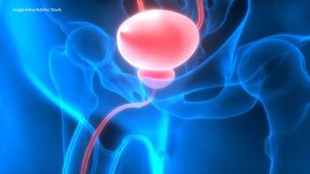
Using 18F-DCPyL PSMA PET/CT can pinpoint more cancers in men who have biochemical failure with initial treatment.

Quantib AI Node is designed for more rapid processing of CT and MRI scans.
Elad Walach, Aidoc chief executive officer, discusses recent research into global CT imaging volume fluctuations and how providers should view that data.
Operational steps are important, but practices who pay attention to broader strategies with patients, insurance, and payment models could see a stronger bounce back.
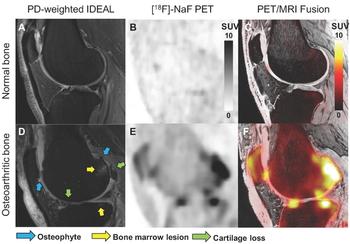
Using PET/MRI 18F NaF offers new functional measure for assessing degeneration of knee joints.

Thoracic vertebrae captured in scans allows for bone mineral density measurements that can pinpoint the presence of osteoporosis.
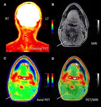
18F-FDG PET/MRI can identify pain generator locations, leading to changes in pain management.
Academic departments share their approaches to COVID-19 recovery.

Residency programs must re-cast the screening and interviewing process in order to be successful during the pandemic.
From February to April 2020, outpatient imaging in rural areas fell 66 percent.
Artificial intelligence tool designed to improve radiologist cancer detection performance.

Paying attention to scanner strength, IV contrast, and body parts imaged can contribute to reducing anesthesia use.
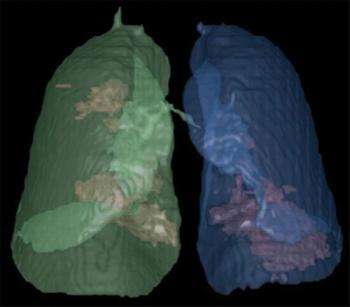
Scans reveal differences in lesion locations, mucoid impaction, and pleural effusion.
Diagnostic Imaging's Weekly Scan; July 10, 2020
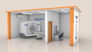
Philips thinks outside-the-box by turning shipping containers into individual CT and X-ray scan rooms.
Case study results show patient has brain connectivity similar to healthy individuals, pointing to positive prognosis possibilities.

An existing program set one London hospital up for success in managing images remotely during COVID-19.