

New ASCO GU data compares fluorinated PSMA PET tracers, showing how bladder activity affects detection of prostate cancer recurrence at low PSA.
New ASCO GU data compares fluorinated PSMA PET tracers, showing how bladder activity affects detection of prostate cancer recurrence at low PSA.
In a recent interview with Diagnostic Imaging, Wendie Berg, MD, PhD, shared her perspective on various aspects of the controversial breast cancer screening guidance issued by the American College of Physicians (ACP), including the use of supplemental imaging in women with dense breasts.
In a recent interview with Diagnostic Imaging, Manisha Bahl, MD, MPH, SBI, offers pertinent insights on the controversial breast cancer screening guidance recently issued by the American College of Physicians (ACP).

Catch up on the top radiology content of the past week.
Real-world PSMA PET use in prostate cancer reveals variable readings and PSA thresholds, prompting selective scans and ongoing trials with FDG PET.
Learn how evolving PSMA PET reporting standards reshape prostate cancer staging and influence therapy choices, from SBRT to triplet regimens.
Learn who reads PSMA PET scans and why a multidisciplinary team translates results into clearer prostate cancer treatment and imaging decisions.
Learn how next‑gen prostate cancer imaging outperforms CT and bone scans, revealing micrometastases and guiding team-based treatment decisions.

In a recent interview, Stamatia Destounis, MD, offered her perspective about the recent American College of Physicians (ACP) guidance on breast cancer screening and concerns over recommendations in women with dense breasts and biennial versus annual mammography screening.

Learn how PSMA PET sharpens staging, guides Pluvicto dosing, and avoids false-positive traps—changing treatment for advanced prostate cancer.
Clinicians weigh PSMA PET’s strengths, limits, and tracer differences, plus when to scan after recurrence to guide salvage prostate cancer therapy.
Learn how FDA-approved PSMA PET tracers differ in prostate cancer staging and recurrence, including urinary uptake nuances that may affect post-prostatectomy detection.
PSMA PET reshapes advanced prostate cancer care, guiding Pluvicto use and tracking response with emerging PCWG4 criteria.
In a recent interview, Matthew Hulse, a certified nuclear medicine technologist, discussed the evolution of amyloid PET imaging in managing patients with Alzheimer’s disease, the impact on PET imaging volume and the emergence of quantification platforms for monitoring treatment efficacy.
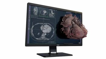
Providing enhanced multidimensional displays of MRI and CT imaging, the Avatar Medical Vision software may facilitate improved surgical planning and intraoperative communication.
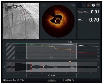
Facilitating enhanced real-time visualization for percutaneous coronary intervention (PCI) procedures, the Ultreon 3.0 optical coherence tomography platform reportedly combines high-resolution imaging with AI-powered insights on stent sizing and positioning.

In recent interviews with Diagnostic Imaging, Wendie Berg, MD, PhD, Stamatia Destounis, MD, and Manisha Bahl, MD, shared their concerns about the recent breast cancer screening guidance from the American College of Physicians.
Catch up on the top radiology content of the past week.
In a recent interview, Manisha Bahl, MD, discussed new breast ultrasound research findings that showed 16 percent higher specificity for breast cancer with adjunctive AI in comparison to unassisted interpretation.

In a recent interview with Diagnostic Imaging, Sujith Kalathiveetil, MD, FACC, discussed the evolution of coronary computed tomography angiography (CCTA), AI-enabled tools and the potential for future innovations with CCTA.
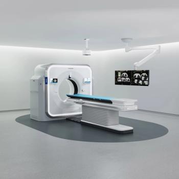
The Rembra CT reportedly facilitates high-throughput imaging for high-volume settings such as emergency departments and the Rembra RT and Areta RT platforms offer an extended 85 cm field of view and 4D CT to enable enhanced accuracy in tumor targeting.

In the latest episode of his “Interventional Radiology in Focus” series, Mina Makary, MD, discusses emerging research and practical pearls for addressing post-thrombotic syndrome in patients with deep vein thrombosis.

In a recent interview, Andrew Del Gaizo, MD, and Nicholas Galante, MD, shared their thoughts and perspectives on increasing imaging volume, the sunsetting of PowerScribe 360, AI hype and promise, and potential opportunities in radiology.
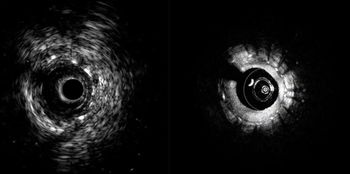
Facilitating real-time visualization of deep vessel structures and high-resolution surface detail within a single workflow, the hybrid intravascular ultrasound (IVUS)/optical coherence tomography (OCT) platform will be launched in the second quarter of 2026.
Catch up on the top radiology content of the past week.
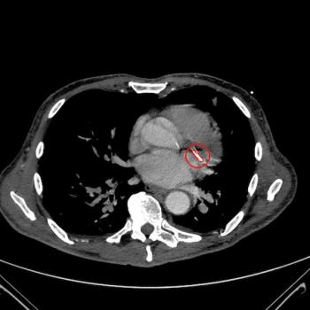
The Bunkerhill Contrast CAC and Bunkerhill Contrast AVC are reportedly the only AI-powered software modalities to garner 510(k) clearance for detection and quantification of coronary artery calcium and aortic valve calcium on contrast-enhanced CT scans.

In a recent interview, Connie Lehman, MD, PhD, discussed key changes to the National Comprehensive Cancer Network (NCCN) guidelines on breast cancer screening, including the use of AI-powered five-year risk assessment beginning at the age of 35.
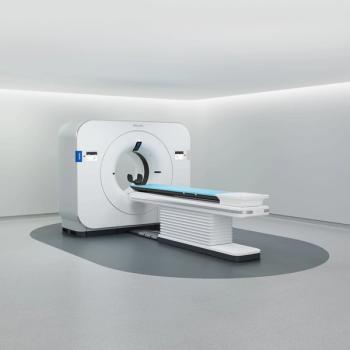
The Philips Spectral CT Verida system reportedly provides reconstruction of 145 images per second, allowing entire CT exams to appear in less than 30 seconds.
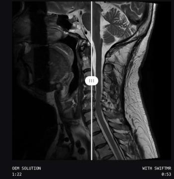
Employing the SwiftMR software with deep learning reconstruction solutions from original equipment manufacturers (OEMs) may provide additional sequence coverage as well as scan time reduction.

In recent interviews with Diagnostic Imaging, Andrew Del Gaizo, MD, Nick Galante, MD and Marc Succi, MD, shared their perspective on the recent comment from NYC Health and Hospitals CEO Mitchell Katz, MD, about potentially replacing radiologists with AI.